
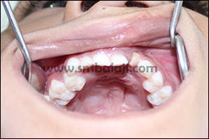
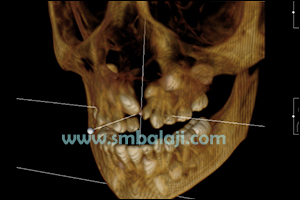
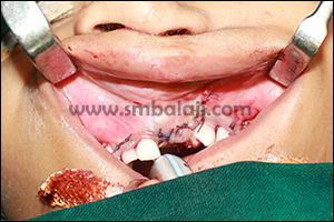
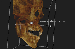
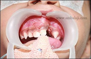
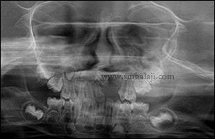
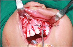
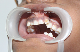
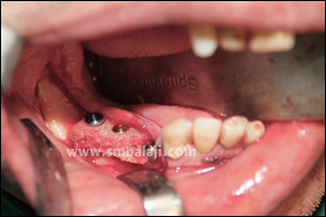
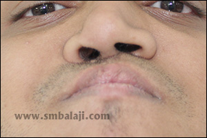
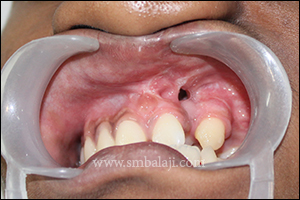
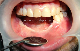
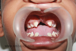
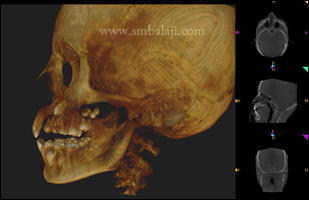
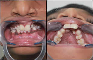
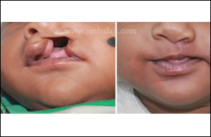
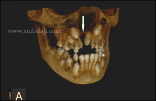
A 6-year-old girl was brought to our hospital by her parents for surgical correction of her cleft defect. Her cleft lip and cleft palate defect was operated elsewhere, in her childhood.
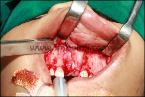
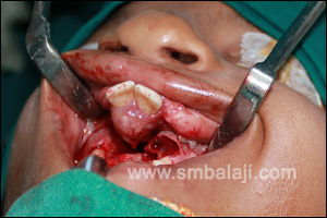
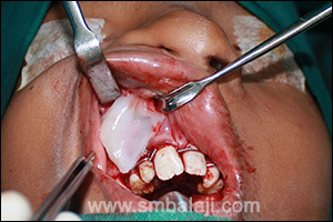
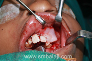
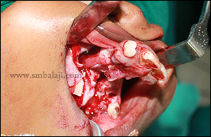
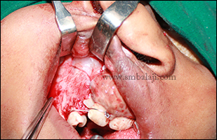
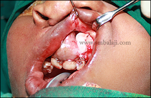
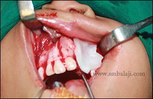
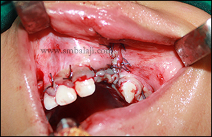
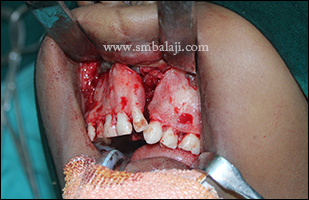
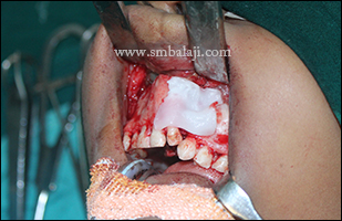
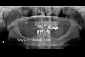
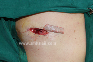
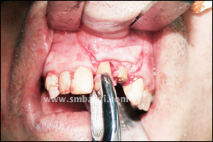
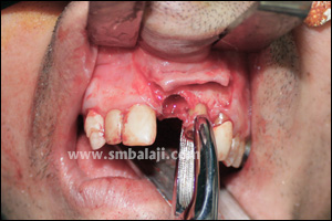
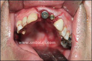
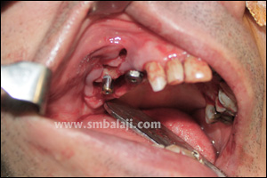
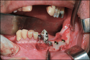
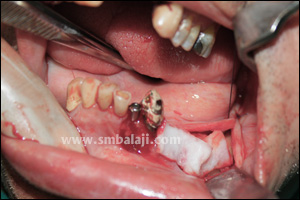
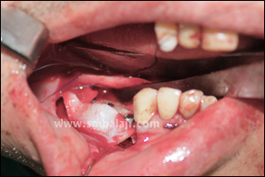
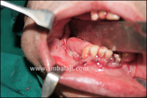
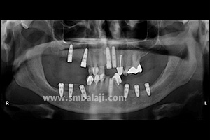
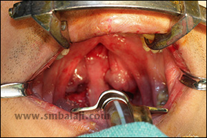
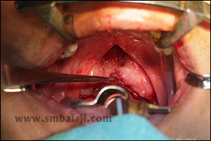
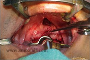
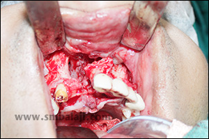
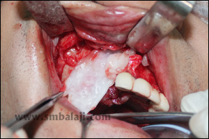
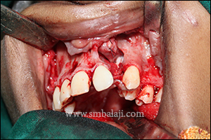
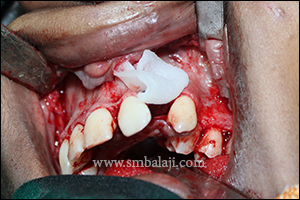
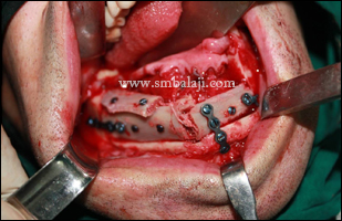
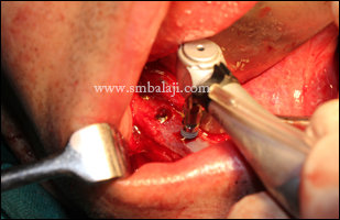
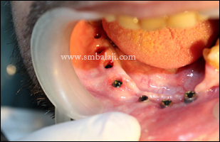
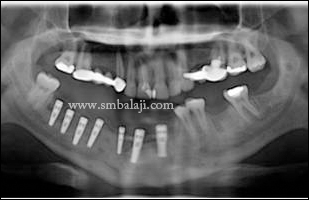
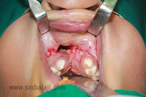
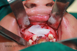
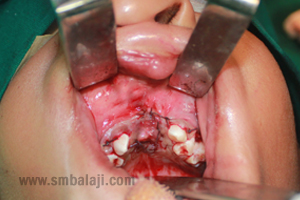
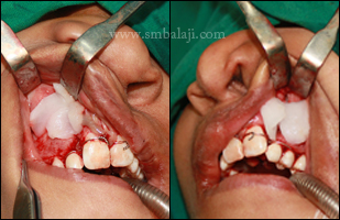
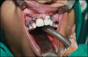
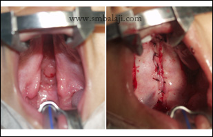
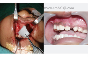
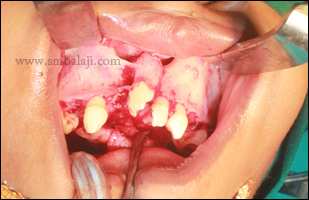
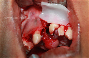
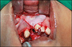
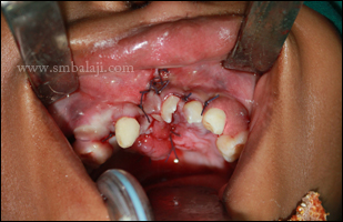
Maxillofacial Surgeon Dr. S.M. Balaji performed the alveolar cleft repair using rhBMP-2 avoiding bone grafting. Miracle protein rhBMP-2 is a bioengineered version of a protein that occurs in the body. When placed in the cleft defect, it stimulates the body’s stem cells to form new bone and thereby healing the bony defect in the teeth bearing region of the upper jaw. By using rhBMP-2, bone graft surgery from the hip or rib is avoided, thereby avoiding an additional surgery & unsightly scars.
Maxillofacial Surgeon Dr. S.M. Balaji performed the alveolar cleft repair using rhBMP-2 avoiding bone grafting. Miracle protein rhBMP-2 is a bioengineered version of a protein that occurs in the body. When placed in the cleft defect, it stimulates the body’s stem cells to form new bone and thereby healing the bony defect in the teeth bearing region of the upper jaw. By using rhBMP-2, bone graft surgery from the hip or rib is avoided, thereby avoiding an additional surgery & unsightly scars.


 RSS Feed