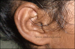
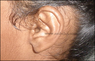
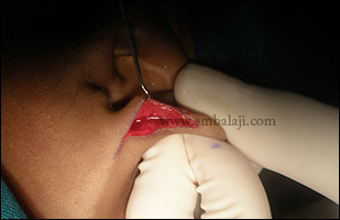
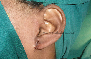
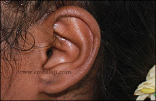
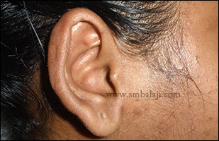
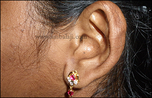
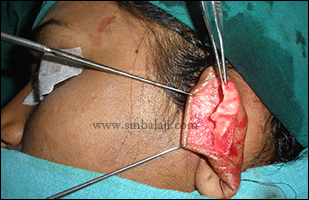
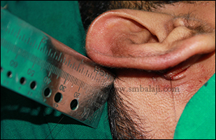
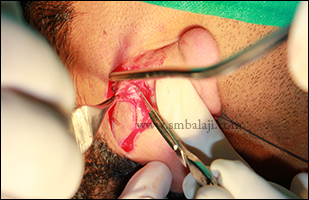
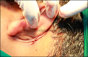
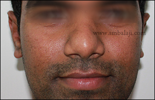
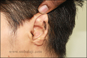
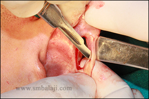
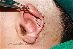
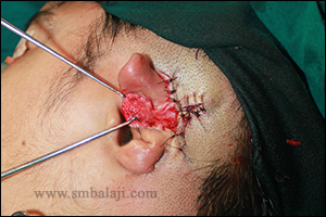
Cosmetic ear correction needs precise skills and that too without any visible scarring. This is a case of left ear lobule detached from the facial skin where the right ear lobule was in correct shape and position. The girl was very keen to wear fashionable earrings but could not do so because the ear lobule shape was different in one ear as compared to the other side. Maxillofacial Surgeon Dr. S. M. Balaji successfully corrected the lobule shape and position without any visible scarring. Thus the patient’s ear shape was made symmetrical as per her wish and she was happy to have the ear correction without any scars on the face.
0 Comments
A 20-year-old girl reported to our hospital seeking to correct the shape of her external ear. Due to excess cartilage the ear helix appeared bulky and enlarged and it appeared asymmetrical. She was not happy with the appearance of the ear shape and wanted to correct it surgically.
Maxillofacial Surgeon Dr. S.M. Balaji successfully performed the ear correction surgery. The excess ear cartilage was surgically trimmed & excess skin tissue was cut. The sutures were placed behind the ear so that there is no visible scar. Immediately following surgery the ear appeared more balanced in shape & size and symmetry was achieved. The girl is very happy to improve her appearance without any scar. A 25 year old man reported to our hospital seeking for the correction of bat ear. Maxillofacial Surgeon Dr. S. M. Balaji successfully corrected his ear shape by removing the skin and excessive cartilage behind the ear using elliptical incision and thus external cartilage of the ear was sutured and well adapted to the skull. Since incision was made behind the ear, no visible scars formed. Results are immediate and the patient is very happy with the surgery outcome. An 18 year old boy was brought to our hospital by his parents for complete reconstruction of his ear defect. He had a large button-holing defect in the helix of his left ear due to which he was teased a lot and it affected his self-confidence. Maxillofacial Surgeon Dr. S.M. Balaji skillfully performed the surgical ear reconstruction in 2 stages. In the first stage the specialized island flap was raised just behind the ear and the edges of the button holing defect in the pinna of the ear was cut. The defective pinna of the ear was sutured with the island flap behind the ear. In the second stage after 6 months, the pinna of the ear was separated from the flap behind the ear and the detached region was sutured. The size of the defect was measured using lint and the required size of skin graft was harvested to close the raw plane of the ventral surface of the ear and it was sutured by means of quilting technique to avoid spacing between the skin graft and recipient site and seroma formation. Thus total reconstruction was done without much surgical scars. Results are immediate and we will update this case by post operative photos with complete healing later. |
Categories
All
Archives
April 2021
|