Best Dental Clinic in Chennai : Best Dental Implant Center in India
FOLLOW US
  • HOME
  • Surgery of The week
  • Clinical Updates
  • Archives
  • Latest News
  • VIDEOS
  • CONTACT US

Dental Implants for Teeth Lost to Gum Disease

6/12/2020

Comments

 
Picture
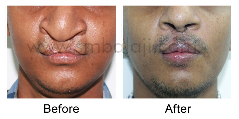
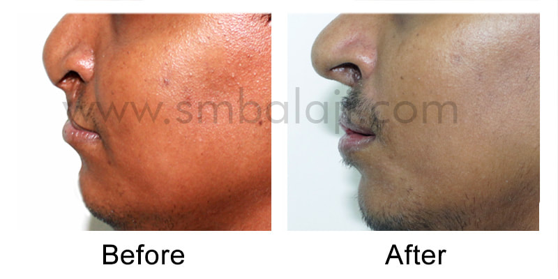
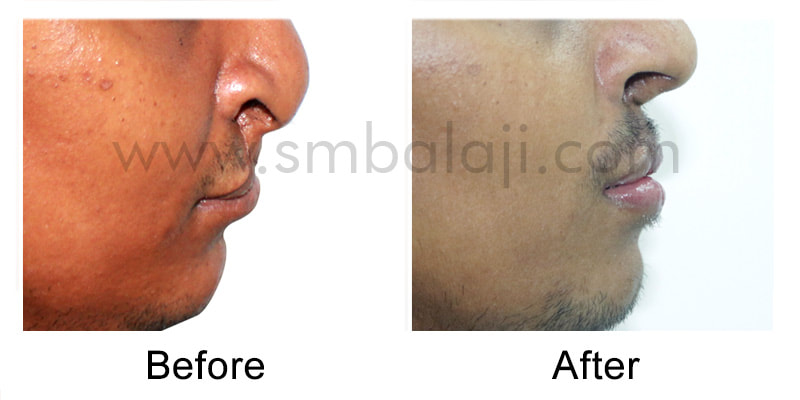
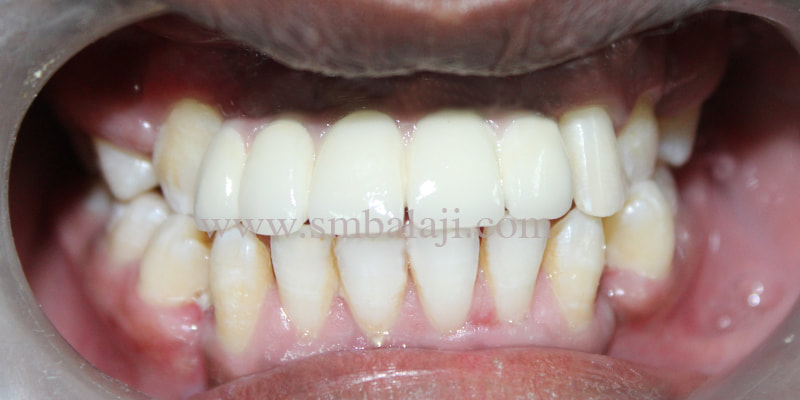
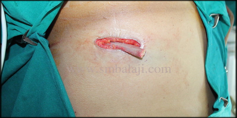
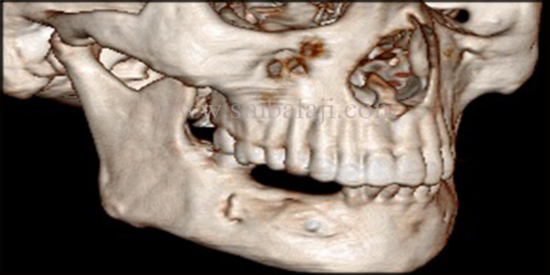
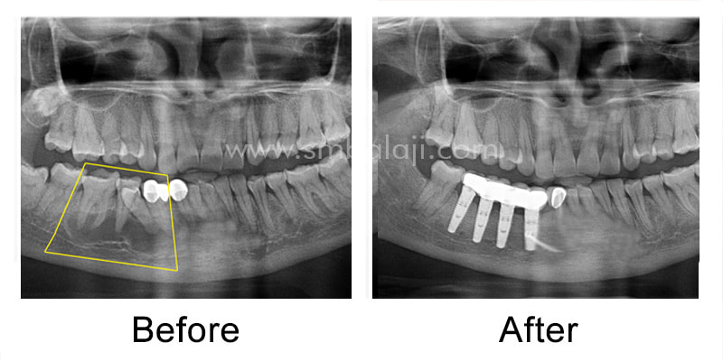
This middle aged man had lost his upper front teeth to a combination of gum disease and diabetes mellitus. Wishing for a permanent solution, he presented to our hospital for dental implants.
​
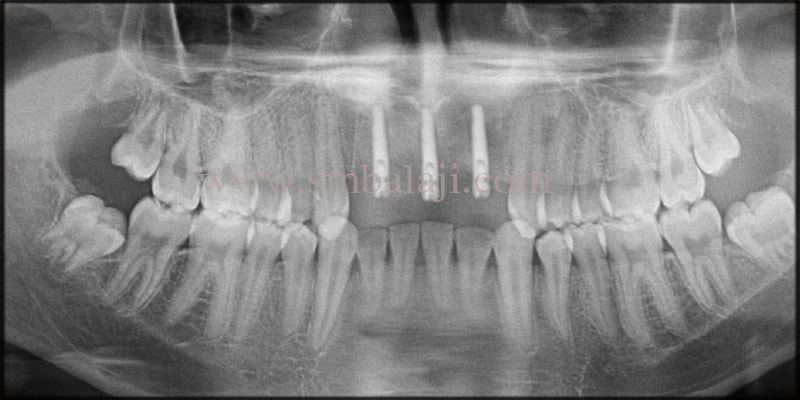
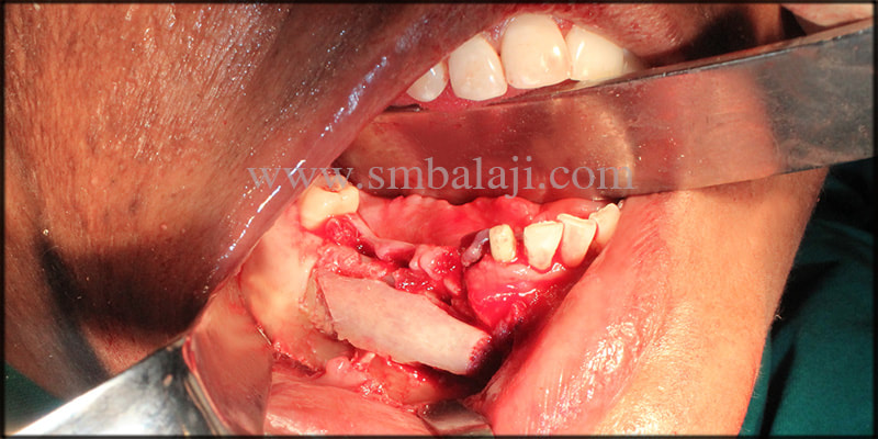
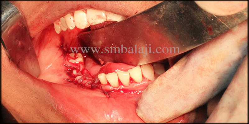
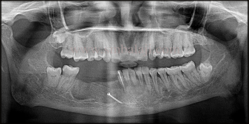
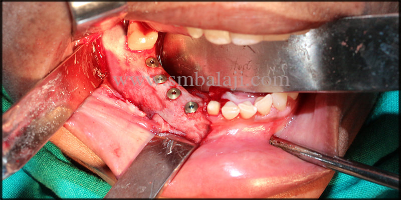
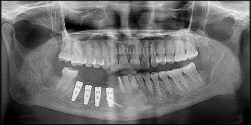
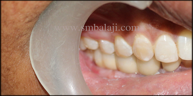
The importance of maintaining meticulous oral hygiene was explained to him. Dental implants were fixed at the site of the missing teeth in his upper jaw. Ceramic crowns were fixed to the dental implants after osseointegration of the implants with surrounding alveolar bone.

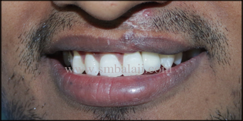
He expressed his happiness at having his teeth back to the hospital staff.
Comments

Dental Implant for Congenitally Missing Lateral Incisor

1/12/2020

Comments

 
This young man had been born with a unilateral cleft lip and palate. His upper left lateral incisor is congenitally missing. He wanted to address this issue as his marriage had been fixed recently and presented to our hospital for dental implant.

A premium dental implant was fixed at the site of the missing tooth followed by a ceramic crown after osseointegration of the dental implant to surrounding alveolar bone.
​
He was extremely happy at the esthetic results of treatment at our hospital.
Comments

Successful Resection of Aneurysmal Bone Cyst Followed by Jaw Reconstruction and Oral Rehabilitation with Dental Implants

15/3/2020

Comments

 
Picture
CASE REPORT

This is a case of a 42 year old female patient who approached our dental hospital with a complaint of asymptomatic swelling in the right lower back teeth region for the past 3 months. The patient stated that the swelling had increased in size over the time. Her medical and family history was unremarkable and there was no history of trauma to the involved site.
The patient was very much in distress due to her condition and wanted to get rid of the cyst.

EXAMINATION OF THE PATIENT

The patient had undergone biopsy of the lesion elsewhere which had diagnosed the swelling to be an aneurysmal bone cyst.
“An Aneurysmal bone cyst (ABC) is a benign, rapidly growing, destructive bone lesion, characterized by replacement of normal bone with blood- filled connective tissue matrix. It is a rare lesion infrequent in craniofacial skeleton”

On extraoral examination, facial asymmetry was apparent with diffuse swelling involving right side of the lower jaw. The swelling was firm and non-tender on palpation. On radiological examination, OPG taken shows the exact extent of the cystic lesion in the right lower jaw extending from the right lower canine tooth till the first molar tooth with expansion of the surrounding bone. On aspiration with a syringe, blood tinged fluid was obtained.

TREATMENT PLAN

Renowned Maxillofacial surgeon, Dr.S.M.Balaji, planned to surgically resect the cystic lesion completely, with extraction of the involved teeth followed by reconstruction of the defective site with rib graft, under general anesthesia. Later replacement of missing teeth with Dental implants was intended.

Dr.S.M.Balaji planned to take rib graft to reconstruct the jaw defect, as Autogenous graft (bone obtained from the same individual receiving the graft ), taken from donor’s extraoral site like chest( rib bone ) has always been regarded to be the “gold standard” due to its properties and absence of immunological reactions. The thick rib graft reduces resorption and helps in bone regeneration.

The patient was clearly explained about the surgical procedure and consent was obtained.
OPG taken shows cystic lesion extending from lower left canine till first molar
Autogenous rib graft taken from the patient’s chest
Cystic lesion removed along with the involved teeth and reconstruction of the defective site with rib graft
Surgical site closed with absorbable suture
Post operative OPG shows well formed bone at the involved site
3DCT scan image shows perfectly formed bone with no bone resorption
Dental implants fixed with stability under local anesthesia
Post operative OPG shows well osseointegrated dental implants with the jaw bone
Perfectly aligned, natural looking fixed ceramic prosthesis
Radiographic image before and after rehabilitation
SURGICAL PROCEDURE
Under general anesthesia, the excision of the lesion was done with complete resection of all tissues lining the cyst and any of its components from the surrounding hard and soft tissues. The involved teeth were also extracted. After successful removal of the cyst, the defective bone area was reconstructed with the rib graft taken from the patient amidst the surgical procedure. The rib graft was fixed with the adjacent jaw bone by means of screws. Finally, the surrounding gum tissues were approximated with absorbable suture.
​
POST SURGICAL FOLLOW-UP

Patient reported after 6 months post surgically for review. Post-operative OPG taken shows well integrated bone graft with the jaw bone. There is no sign of recurrence of the cyst. A 3DCT scan taken revealed perfectly formed bone at the involved site with no signs of bone resorption, which took us to the final step of replacing her missing teeth with dental implants.

DENTAL IMPLANT PLACEMENT

Under local anesthesia, Dr.S.M.Balaji opened the gum tissues surrounding the relative site exposing the newly formed bone. Dental implants of appropriate size and height were fixed in the underlying jaw bone with precision. Finally the gum tissue were closed with absorbable suture.
The patient was asked to wait for a period of 3-4 months for the final prosthesis, as to allow the dental implants to completely osseointegrate with the jaw bone obtaining maximum stability and retention. Meanwhile, a removable prosthesis was given temporarily for replacement of missing teeth to avoid supraeruption of the opposing teeth.
 
THE HAPPY PATIENT

The patient returned after 4 months for the final prosthesis. OPG taken shows dental implants in integration with the jaw bone. Hence, the final impression was taken and after a couple of bite trials, a natural looking fixed ceramic bridge was fixed onto the dental implants. The prosthesis was in perfect contact with the opposing teeth. The patient and her family members were very happy and satisfied with the entire process and the outcome. Patient was able to chew properly without any discomfort. Patient is on a regular follow up for the past 2 years.
Comments

    Categories

    All
    Abbe Flap
    Apicectomy
    Bone Grafting
    Buccal Mucosa Lesion
    Ceramic
    Ceramic Prosthesis
    Cosmetic Dentistry
    Cosmetic Surgery
    Crown
    Dental Implant
    Excision Biopsy
    Hairy Nevus Removal
    Immediate Implant
    Implant
    Jaw Reconstruction
    Operculectomy
    Orthodontics
    Periodontal
    Prosthesis
    Rehabilitation
    Root Canal (RCT)
    Sinus- Lift
    Smile Makeover
    Teeth Whitening
    Tooth Extraction
    Tooth Restoration
    Zygoma Implant

    Archives

    January 2021
    December 2020
    March 2020
    January 2018
    February 2017
    August 2016
    July 2016
    May 2016
    April 2016
    February 2016
    January 2016
    February 2015
    January 2015
    November 2014
    September 2014
    August 2014
    July 2014
    June 2014
    May 2014
    April 2014
    March 2014
    February 2014
    December 2013

    RSS Feed

Copyright © 2020 Balaji Dental & Craniofacial Hospital | All rights reserved | Powered by BALAJI DENTAL HOSPITAL