Best Dental Clinic in Chennai : Best Dental Implant Center in India
FOLLOW US
  • HOME
  • Surgery of The week
  • Clinical Updates
  • Archives
  • Latest News
  • VIDEOS
  • CONTACT US

Operculectomy & orthodontics to align impacted canine tooth

22/6/2014

Comments

 
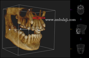
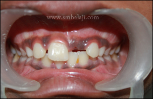
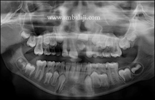
A 15-year-old boy was brought to our dental clinic by his parents seeking to align his crookedly placed teeth. His upper right canine tooth had not erupted. A specialized 3D CBCT scan taken showed that the canine tooth was impacted i.e abnormally positioned high above near the roots of adjacent teeth.

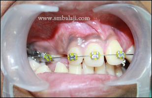
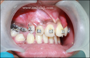
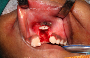
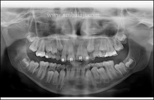
The canine teeth are important in the dental arch as they frame the smile and play a critical role in biting and chewing. To bring the impacted tooth into proper alignment, an operculectomy was done. This is a surgical procedure in which the gum on top of the impacted tooth was removed to expose the hidden tooth underneath. Once the tooth was exposed, an orthodontic bracket was bonded to the exposed tooth. With subsequent orthodontic treatment, the tooth was brought into normal position maintaining its vitality and the teeth were aligned to improve function & appearance. 
Missing upper canine tooth in the right side
3D CBCT scan image showing buccally impacted canine tooth
After operculectomy and subsequent orthodontic treatment canine tooth was brought into occlusion
Comments

Successful Operculectomy & Unerupted tooth brought to its normal position through orthodontics

18/3/2014

Comments

 
A 10 year-old boy was brought to our hospital by his parents with a complaint of an unerupted upper front tooth. The front tooth on the right side had erupted normally but the tooth on the left side had not appeared.

A digital X-ray taken showed an over-retained deciduous tooth (milk tooth) preventing the normal eruption of the permanent maxillary left central incisor. The deciduous tooth was extracted and with the operculectomy procedure the thick overlying soft tissues were uncovered to expose the “hidden” permanent tooth. Using an orthodontic appliance, the tooth was brought down into normal position maintaining its vitality. This was performed at an earlier stage itself to prevent the impaction of central incisor. When all the milk teeth shed, orthodontic treatment will be done to align the teeth.
Preoperative intraoral view showing missing permanent upper left central incisor
Preoperative Xray showing retained impacted deciduous and upper central incisor
Impacted retained deciduous tooth removed surgically
Impacted upper permanent incisor exposed surgically
Postoperative Xray showing upper incisors brought to alignment
Postoperative clinical view with braces
Comments

    Categories

    All
    Abbe Flap
    Apicectomy
    Bone Grafting
    Buccal Mucosa Lesion
    Ceramic
    Ceramic Prosthesis
    Cosmetic Dentistry
    Cosmetic Surgery
    Crown
    Dental Implant
    Excision Biopsy
    Hairy Nevus Removal
    Immediate Implant
    Implant
    Jaw Reconstruction
    Operculectomy
    Orthodontics
    Periodontal
    Prosthesis
    Rehabilitation
    Root Canal (RCT)
    Sinus- Lift
    Smile Makeover
    Teeth Whitening
    Tooth Extraction
    Tooth Restoration
    Zygoma Implant

    Archives

    January 2021
    December 2020
    March 2020
    January 2018
    February 2017
    August 2016
    July 2016
    May 2016
    April 2016
    February 2016
    January 2016
    February 2015
    January 2015
    November 2014
    September 2014
    August 2014
    July 2014
    June 2014
    May 2014
    April 2014
    March 2014
    February 2014
    December 2013

    RSS Feed

Copyright © 2020 Balaji Dental & Craniofacial Hospital | All rights reserved | Powered by BALAJI DENTAL HOSPITAL