Best Dental Clinic in Chennai : Best Dental Implant Center in India
FOLLOW US
  • HOME
  • Surgery of The week
  • Clinical Updates
  • Archives
  • Latest News
  • VIDEOS
  • CONTACT US

Successful primary lip repair for unilateral cleft lip and palate baby boy

28/2/2015

Comments

 
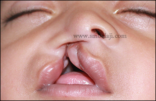
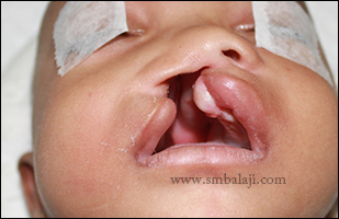
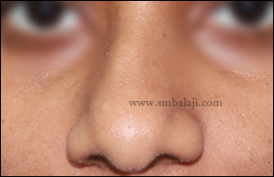
A 3-month-old baby boy born with unilateral cleft lip and palate was brought to our hospital by his parents for specialized treatment of the defect.

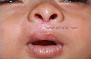
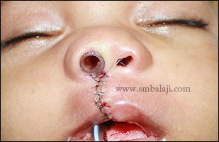
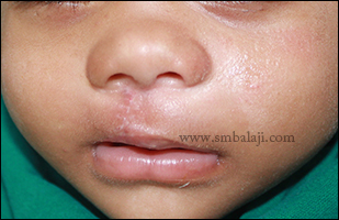
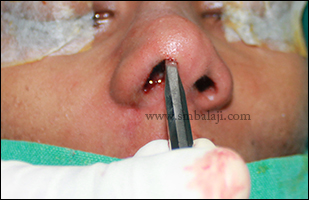
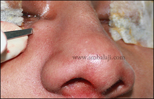
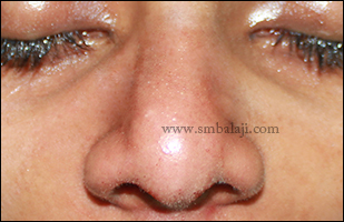
Maxillofacial Surgeon Dr. S.M. Balaji successfully performed the primary cleft lip repair using modified Millard’s technique.  Results are immediate. The baby’s appearance was remarkably improved and was able to feed well. His parents were very happy to have the cleft lip surgically treated with negligible scar. After few months cleft palate repair will be done.
Unilateral cleft lip and palate in 3 months old baby boy
Immediately after suturing the cleft lip defect
Enhanced appearance of the baby immediately after suture removal following seven days of surgery
Comments

Successful jaw reconstruction & dental implants

25/2/2015

Comments

 
A young man from Middle East Asia suffering from Ectodermal Dysplasia was referred to our Hospital for expert care. He had complete missing teeth (anodontia) from childhood. Due to this there was progressive loss of alveolar process. He had consulted many dental surgeon for the same but due to reduced width and height of the alveolar process they refused to give dental  implant replacement for his missing teeth.  

Maxillofacial Surgeon and Dental Implantologist Prof. S.M Balaji performed the complete jaw bone reconstruction using rib graft & rhBMP-2. Implants were placed to replace his missing teeth in mandible. Following osseointegration of the implants, ceramic prosthesis will be given. Due to very thin alveolar process in maxilla, costochondral rib graft and BMP placed to enhance new bone formation. Later dental implants with good retention and stability will be placed after one month.
Intraoral view showing completely edentulous maxilla and mandible
3DCT scan view showing complete anodontia of both maxilla and mandible with thin alveolar process
Costochondral rib graft harvested to reconstruct the receded portion of alveolar process
Dental implants fixed in the lower jaw and stabilized with rib graft for good retention
Harvested rib graft along with miracle protein rhBMP2 used to reconstruct the alveolar process
Immediately after suturing following complete reconstruction of maxilla and mandible
Postoperative digital orthopantomogram showing graft stabilization and good dental implant retention in mandible
Comments

Successful removal of large oral tumor and reconstruction without any scars

23/2/2015

Comments

 
This is a case of adenomatoid odontogenic tumor involving maxilla and zygoma on the right side. Patient came from West Bengal with the complaints of huge swelling in the right side of the face. The swelling was progressive and initially he had pain in his right upper jaw and cheek. He had consulted many doctors for removal of this huge swelling without any scars but they were refused to do intraorally. One of his friends referred him to us for the treatment.

After thorough clinical, radiological and histopathological examination, Maxillofacial Surgeon Dr. S. M. Balaji successfully removed the entire tumor mass intraorally along with sufficient clearance to avoid recurrence. Following surgery his facial appearance was improved dramatically and after a week satisfactory healing was achieved intraorally. Patient feels very happy to have the entire correction done in a single surgery and done without any visible surgical marks on his face.

Preoperative view showing swelling in the right side of the face obliterating the nasolabial fold
Note the elevation of the ala of nose on the right side to to huge swelling
Note the localized bluish discoloration of the swelling suggesting of the cystic change
Preoperative 3DCT scan view showing complete destruction of bone due to odontogenic tumour on the right side involving maxilla and zygoma
Intraoperative view showing complete removal of the lesion along with the affected bone intraorally
Complete excision of the tumor mass in toto
Immediately after suturing
Postoperative intraoral view following 10 days from surgery showing satisfactory healing
Enhanced appearance following surgery without any scars on the face
Comments

Surgical treatment for Oral Submucous Fibrosis

20/2/2015

Comments

 
A 30-year-old man came to our hospital seeking treatment for his inability to open his mouth normally. Preoperatively, his mouth opening was just 11 mm. He gave a history of cigarette smoking and pan chewing for 2 decades. He was diagnosed with Oral submucous fibrosis, a condition characterized by thickening of the mucosal tissues and progressive rigidity of the jaws making it exceedingly difficult to open the mouth.

In the surgical treatment procedure, Maxillofacial Surgeon Dr. S.M. Balaji placed transverse incisions on the cheek on both right & left side to release the fibrous bands and to immediately improve the mouth opening. An inferiorly based Nasolabial island flap based on the facial artery was raised and transposed intraorally through a small trans-buccal tunnel. This was sutured with the buccal mucosal defect. Extraorally, the defect was closed in layers. Immediately following surgery, a normal mouth opening of 32 mm was achieved.
Preoperative reduced mouth opening
Nasolabial flap raised to reconstruct the affected portion of buccal mucosa
Nasolabial flap rotated and fixed in the defective portion of buccal mucosa
Immediately after surgery
Increased mouth opening following surgery
Comments

Successful cosmetic cleft nose correction with lip revision

18/2/2015

Comments

 
A 28-year-old man from Kolkata reported to our hospital seeking specialized treatment to correct his cleft nose defect. He was previously operated for cleft lip and palate elsewhere in his childhood. Due to the cleft defect his nose was collapsed on the right side and it affected his facial appearance. He wanted to have unsightly lip scar correction also in the same surgery.

Maxillofacial Surgeon Dr. S.M. Balaji successfully performed the rhinoplasty or nose correction. A costochondral graft (rib graft) was harvested and used to reconstruct the collapsed nasal framework. The surgery was approached from inside the nose so there will be no scar formation. Alar base on the right side was at a lower level compared to other side due to cleft defect. This was corrected by raising the floor of nose intraorally using costochondral graft thus nose was made symmetrical on either side. 

Huge unsightly lip scar defect which was formed due to previous lip surgery done elsewhere also corrected and shape of the vermillion was reconstructed. Immediately after surgery, the nose appeared sharp and definite giving a more pleasing appearance. The patient was very happy to have his nose and lip defect corrected without much visible surgical marks. 
Preoperative facial view showing asymmetric nose profile and depressed alar base on the left side due to cleft defect
Preoperative view showing depressed alar base on the left side due to cleft bone defect in the base of the nose
Harvested costochondral rib graft to augment the defective cleft nose
Costochondral rib graft used to raise the depressed dorsum of the nose
Cleft lip revision and augmented dorsum of the nose giving enhanced appearance
Alar base on the left side of the nose was raised and approximated to the right side without any visible surgical marks
Comments

Complete facial rehabilitation in a complex facial bone fracture

16/2/2015

Comments

 
This is a case of multiple facial bone fracture due to road traffic vehicular accident happened 5 months back. The patient had first aid treatment and fractured bone stabilization at nearby hospital elsewhere. But he was not happy with the surgical outcome where left eye was at a lower level compared to the right side. He was not able to bite properly which made him very uncomfortable.

On examination, facial appearance was completely changed and he was not able to bite properly due to improperly united maxillary and Mandibular fracture segments. Left Mandibular canine and lateral incisor teeth were fractured and canine crown was displaced in between the fracture margins of the mandible which was not removed in the previous surgery done elsewhere.

After thorough clinical and radiological examination, Maxillofacial surgeon Dr. S. M. Balaji diagnosed that his left lateral orbital rim and zygomatic arch were fractured and laterally displaced. Maxilla and mandible also were fractured and wrongly united which led to improper bite. Dr. Balaji planned to correct his facial appearance and bite in a single surgery. Intraorally the impacted canine crown was removed along with the fractured lateral incisor and replaced with dental implants. Both upper and lower jaw stabilized and fixed in proper occlusion. Lateral orbital rim was also brought medially by intraocular incision and floor of the orbit was raised using titanium mesh. Thus the fractured facial bones were corrected in such a way that gave an improved appearance without any visible scars on his face. 
Preoperative view showing depressed left orbital content
3DCT scan view showing multiple facial bone fractures stabilized with bone plates
Malunited fracture margins of mandible and displaced canine crown into the fractured segments
Displaced canine crown was exposed intraorally and the fracture segments were approximated
Canine crown was removed in toto
Fractured left lower lateral incisor and canine teeth were extracted and dental implants fixed into the sockets
Displaced portion of left lateral orbital rim was approximated into its correct position
Entire orbital content was raised and made symmetrical with that of the right side using titanium mesh.
Immediate postoperative view showing raised left eye and made symmetrical without any damage to the orbital content
Comments

Cosmetic correction of misaligned left eyebrow due to a trauma treatment done elsewhere

14/2/2015

Comments

 
A 22 year old man reported to our hospital seeking cosmetic correction of his left eyebrow. One year back, due to an accident he had a cut injury in the left eyebrow for which he had a first aid at some hospital elsewhere. The cut ends of the eyebrow were misaligned which made him very conscious about his facial appearance. And also he wanted to correct the scar which was wide and unsightly formed due to previous surgery.

Maxillofacial Surgeon Dr. S. M. Balaji successfully corrected the defect as well as the wide scar formed due to previous surgery. Dr. Balaji approximated the misaligned ends of the eyebrow and corrected the unsightly scar by removing the thick scar tissue. Patient is happy to have cosmetic correction of his eyebrow which enhanced appearance significantly. 
Splitted and misaligned left eyebrow due to previous surgery done elsewhere
Immediate postoperative view where the eyebrow was corrected and the ends were sutured to its correct position
Immediately after suture removal following seven days from surgery showing enhanced appearance
Comments

Primary lip repair for unilateral cleft lip & palate

13/2/2015

Comments

 
A 3-month old baby boy born with unilateral cleft lip & palate was brought to our hospital by his parents seeking the best treatment for cleft defect.

Maxillofacial Surgeon Dr. S.M. Balaji performed the primary lip repair surgery using Modified Millard’s technique. Following surgery, the baby’s appearance improved greatly and he was able to feed well. The parents were very happy that their baby had hardly any post-surgical scars. Consecutively cleft palate correction surgery will be done.


Unilateral cleft lip and palate in a 3 months old baby boy
Primary cleft lip closure using modified Millard's technique
Immediately after suture removal following seven days of surgery
Comments

Deviated nose correction without any scars

11/2/2015

Comments

 
A 21-year-old man reported to our hospital seeking to correct his deviated nose which greatly affected his facial appearance. He was very much disappointed as his nose was deviated to his left side which was very obvious in photographs.

Maxillofacial Surgeon Dr. S.M. Balaji successfully performed the closed rhinoplasty technique. Deviated nasal septum was corrected by lateral osteotomy technique and resection of caudal portion of nasal septal cartilage on the right side. Thus entire surgery was done from inside the nose to avoid any scars on the face (closed rhinoplasty technique).

A balance between the nose shape and facial profile was achieved for a more pleasing appearance. The patient was very happy to have a corrected nose that enhanced his facial profile with absolutely no scarring.  

Preoperative view showing deviated nose towards left side with prominent lower lateral nasal septal cartilage
Comments

Cosmetic correction of frontal bossing without any scars

9/2/2015

Comments

 
A 25 year old man from Bangladesh reported to our hospital seeking cosmetic correction of his forehead bossing. He had this prominence from his childhood itself. He was very upset of his profile view where the forehead prominence was very obvious. He wanted to reduce this cosmetically without any scars.

After thorough clinical and radiological examination, Maxillofacial Surgeon Dr. S. M. Balaji diagnosed that due to frontal sinus enlargement the frontal bone was bulged out. Dr. Balaji corrected this defect by raising bicoronal flap and the outer table of frontal bone prominence was reduced. The frontal sinus cavity was explored and to maintain the forehead flatness titanium mesh was placed.  Results are immediate and the patient is happy to have entire correction done without any scars on the face.
Sagittal view showing increased frontal sinus space leading to bossing
Facial profile view showing prominent frontal bossing
Intraoperative view where prominent frontal plate was exposed
After reducing the frontal bone excess to maintain the shape of the forehead the frontal sinus cavity was raised with titanium mesh
Postoperative profile view showing flat forehead giving enhanced appearance without any scars
Comments

Secondary cleft palate with bifid uvula correction

6/2/2015

Comments

 
This was a case of isolated cleft palate where primary palate repair was done elsewhere. The patient came to our hospital with the complaints of escape of water through the nose due to small hole in the mid palatine region.

After thorough clinical examination, Maxillofacial Surgeon Dr. S. M. Balaji diagnosed it as oronasal fistula in the mid palatine region and bifid uvula due to post surgical defect after treatment done elsewhere. By following palatal pushback technique, re palate was done and bifid uvula was joined. Results are immediate and positive suction test is achieved. 
Preoperative view showing bifid uvula
Failed palate repair having oronasal fistula
Oronasal fistula closed using palatal pushback technique and Bifid uvula corrected
Video
Comments

Cosmetic rhinoplasty for devaited nose without scars

4/2/2015

Comments

 
A 22 year old woman reported to our hospital seeking to correct her deviated nose which greatly affected her facial appearance. She was very much disappointed as her nose was completely deviated to the right side giving asymmetric facial appearance.

Maxillofacial Surgeon Dr. S.M. Balaji skillfully performed the cosmetic rhinoplasty. Vertically nasal septal cartilage height was trimmed by medial osteotomy to reduce the height of the nose. Width of the nose was also reduced by lateral osteotomy technique. Caudal portion of lateral nasal septal cartilage was removed to correct the nose tip. Thus deviated nose and size of the framework of the nose was corrected from inside the nose to avoid any scars on the face (closed rhinoplasty technique).

A balance between the nose shape and facial profile was achieved for a more pleasing appearance. The patient was very happy to have a narrower, shapely nose that enhanced her facial profile with absolutely no scarring. 
Preoperative view - deviated nose towards right side
During procedure - lower lateral nasal septal cartilage removed intranasally
Medial osteotomy of the nose to reduce the height of the nose
Lateral osteotomy to correct the deviated nasal bridge
Immediate postoperative view showing straightened nasal septum giving pristine look without any visible surgical marks
Comments

    Categories

    All
    Abbe Flap Technique
    Advanced Transport Distraction Osteogenesis
    Advancement Genioplasty
    Alveolar Cleft Repair
    Alveolar Distraction
    Ameloblastoma
    Augmentation Genioplasty
    Augmentation Rhinoplasty
    Bat Ear Correction
    Bilateral Cleft Lip Palate
    Blepharoplasty
    Bone Defect Reconstruction
    Bone Grafting
    Bone Morphogenetic Protein(BMP)
    Broad Flattened Nose Correction
    Bulky Nose Correction
    Chin Correction
    Chin Correction Surgery
    Cleft Lip & Palate Surgery
    Cleft Nose
    Cleft Palate Repair
    Cleft Rhinoplasty
    Complete Facial Rehabilitation
    Complex Facial Fractures
    Condylar Fracture Surgery
    Correction Of Hypoplastic Ramus
    Corrective Jaw Surgery
    Cosmetic Cleft Nose Correction
    Cosmetic Nose Correction
    Cosmetic Rhinoplasty
    Cosmetic Surgery
    Craniofacial Deformities
    Craniofacial Reconstruction
    Craniofacial Surgery
    Cyst Removal
    Dental Implant
    Dentigerous Cyst
    Deviated Nose Correction
    Diplopia
    Distraction Osteogenesis
    Ear Correction
    Ear Defect
    Ear Reconstructive Surgery
    Epiphora
    Extruding Infected MEDPOR
    Eyebrow Correction
    Face Enhancement
    Face Reconstruction
    Facial Asymmetry Correction
    Facial Bone Fracture
    Facial Burn Injury
    Facial Palsy Surgery
    Fibrous Dysplasia
    Flat Nose Correction
    Forehead Bone Fracture
    Forehead Deformity
    Forehead Flap
    Frontal Bossing Correction
    Gummy Smile
    Hemifacial Microsomia
    Implant
    Incomplete Cleft Lip
    Isolated Cleft Palate Repair
    Jaw Correction Surgery
    Jaw Cyst
    Jaw Deformity
    Jaw Fracture Surgery
    Jaw Joint Ankylosis
    Jaw Reconstructive Surgery
    Jaw Surgery
    Lip Defect Correction
    Lip Revision
    Locked Jaw
    Lower Jaw Fracture
    Low Lying Upper Eyelid Correction
    Macrostomia Correction
    Mandible Correction
    Mandible Reconstruction
    Maxillofacial Surgery India
    Maxillomandibular Distraction
    Maxilofacial Surgery
    Micrognathic Mandible Correction
    Microtia
    Nasal Glial Heterotopia
    Naso-orbito-ethmoidal Fracture
    Nose And Lip Correction
    Nose Asymmetry Correction
    Nose Correction
    Nose Job
    Nose Reconstructive Surgery
    Oral Submucous Fibrosis
    Oral Tumor Removal
    Orbital Correction
    Orehead Defect
    Orthognathic Surgery
    Parrot-beak Nose
    Parry Romberg Syndrome
    Pharyngoplasty
    Plastic Surgery
    Primary Cleft Lip Repair
    Profile EnhancemeP
    Reduction Glossectomy
    Reduction Rhinoplasty
    Removal Of Capillary Hemangioma
    RhBMP 2
    RhBMP-2
    Rhinoplasty
    Road Accident Trauma
    Road Accident - Trauma
    Scar Revision
    Secondary Cleft Palate
    Sinus Lift
    Smile Makeover
    Speech Correction
    Square-face Correction
    Surgery Without Scar
    Tessier Nose Defect
    Tissue Expansion
    TMJ Ankylosis
    Tongue Correction
    Tongue Surgery
    Trauma
    Trauma Treatment
    Triangular Shaped Forehead
    Tumor Resection
    Unilateral Cleft Lip
    Unilateral Cleft Lip And Palate
    Unilateral Cleft Lip Repair
    Velopharyngeal Incompetence

    Archives

    April 2020
    March 2019
    February 2019
    January 2018
    November 2017
    October 2017
    September 2017
    June 2017
    May 2017
    April 2017
    March 2017
    January 2017
    December 2016
    October 2016
    September 2016
    August 2016
    July 2016
    June 2016
    May 2016
    April 2016
    March 2016
    February 2016
    January 2016
    December 2015
    October 2015
    September 2015
    August 2015
    July 2015
    June 2015
    May 2015
    April 2015
    March 2015
    February 2015
    January 2015
    December 2014
    November 2014
    October 2014
    September 2014
    August 2014
    July 2014
    June 2014
    May 2014
    April 2014
    March 2014
    February 2014
    January 2014
    December 2013
    November 2013

    RSS Feed

Copyright © 2020 Balaji Dental & Craniofacial Hospital | All rights reserved | Powered by BALAJI DENTAL HOSPITAL