Best Dental Clinic in Chennai : Best Dental Implant Center in India
FOLLOW US
  • HOME
  • Surgery of The week
  • Clinical Updates
  • Archives
  • Latest News
  • VIDEOS
  • CONTACT US

Successful mandible reconstruction with recombinant technology for failed fibular graft

31/10/2014

Comments

 
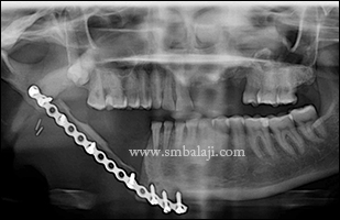
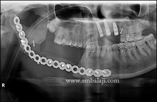
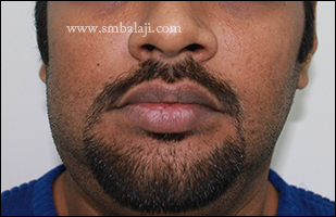
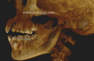
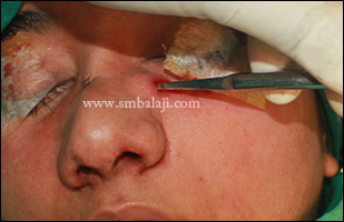
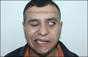
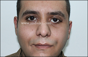
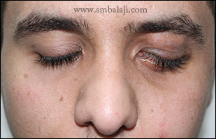
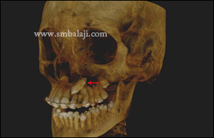
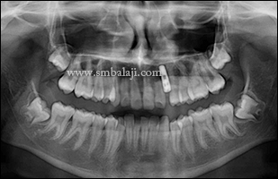
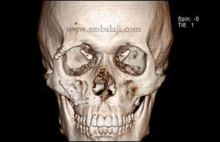
A 46 year old female patient reported to our hospital for swelling and pus drainage from right side of mandible.  She had sustained a road traffic accident 3 years ago and had undergone multiple surgeries for the same. Her reports and x-rays showed that she had undergone free fibular graft treatment elsewhere. Currently, there was an associated swelling and pus drain from the region.  On examination,   there was graft dehiscence in the retromolar region with draining extraoral sinus.   The CT scan of the patient revealed a fibula graft and incorrectly contoured straight reconstruction plate.

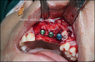
Maxillofacial Surgeon Dr. S. M. Balaji successfully planned to surgically remove the infected graft and place a composite mandibular reconstruction with rib graft fixed with reconstruction plate and reinforced with rhBMP-2. As she also had a missing 21, 22, 23, dental implants were also planned as a part of this procedure.

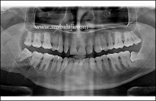
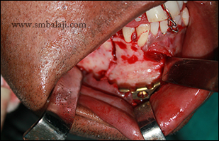
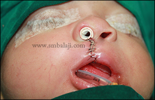
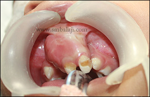
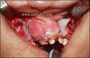
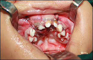
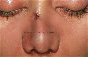
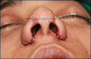
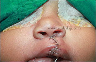
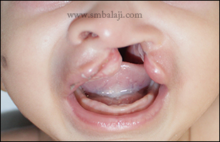
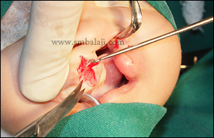
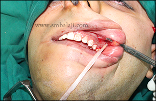
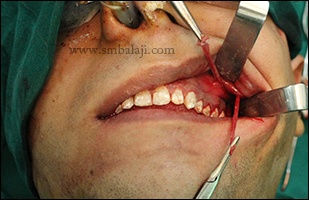
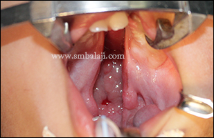
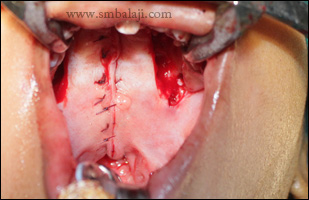
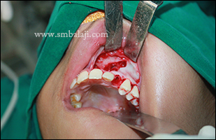
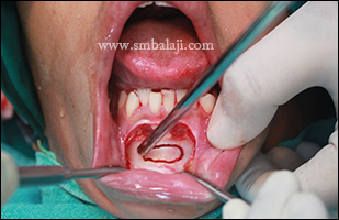
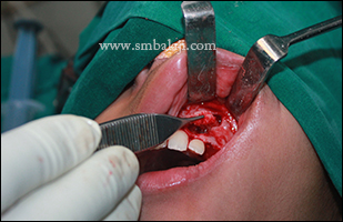
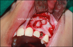
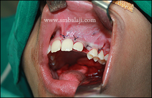
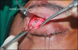
Intraorally, crestal incision was placed from tooth 21 to 23 regions. Mucoperiosteal flap reflected, implants placed and flap repositioned and closed. Later after osseo-integration, super structures would be placed.

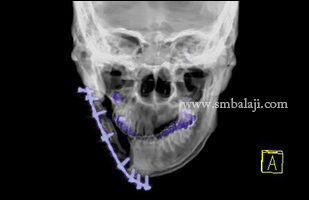
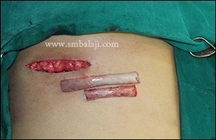
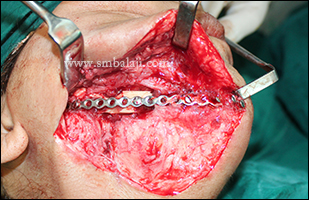
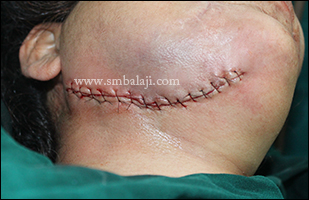
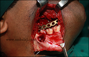
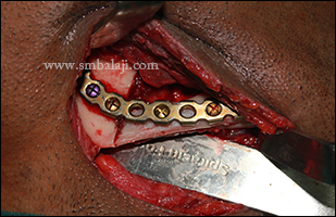
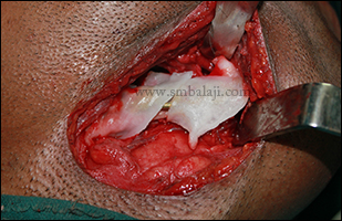
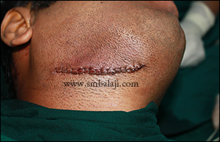
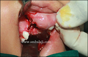
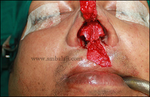
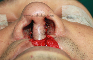
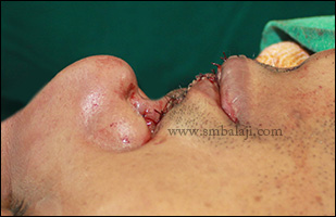
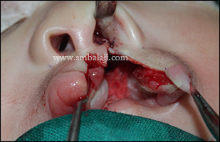
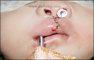
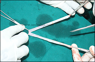
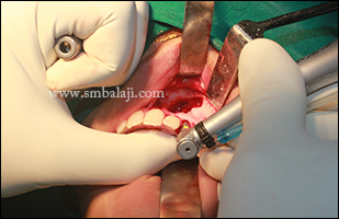
Right submandibular incision was placed through the existing scar. The scar and the sinus tract were carefully excised. The reconstruction plate along with resorbed fibular graft was identified and removed. A new titanium reconstruction plate was contoured, adapted and fixed to the jaw bone using screws. The rib graft was now placed along the medial aspect of the reconstruction plate and stabilized using screws. rhBMP-2 was placed over this reconstructed area and the closure was done layer wise.  rhBMP-2 will induce new bone formation that will ensure complete healing of the defect.

Preoperative orthopantamogram showing infected plate with failed fibular graft on right side of the lower jaw
3D CT scan of the lower jaw on the right side showing failed bone graft with defective bone formation
Upper missing teeth were replaced with dental implants with defective alveolar bone region enhanced with bone graft
Costal graft harvested for reconstruction of the right side of the mandible
Submandibular incision made to expose the infected plate with failed fibular graft
Infected bone graft with the bone plate along with bone screws were removed
Harvested costal graft stabilized with long titanium bone plate used to reconstruct the lower jaw
Miracle protein rhBMP-2 was wrapped over the costal graft to induce new bone formation
Submandibular incision was sutured by layer closure
Immediate postoperative orthopantomogram showing complete removal of infected bone and reconstruction of mandible with costal graft
Comments

Jaw Reconstruction with recombinant technology for tumor resection

29/10/2014

Comments

 
A 32 year old man reported to our hospital with the complaint of swelling in the right side of his face. He added that the size of the swelling was increasing progressively which made his face asymmetrical. He was very cautious about this progressive swelling which made him under huge depression.

After thorough clinical and radiological examination, Maxillofacial Surgeon  Dr. S. M. Balaji diagnosed it as ameloblastoma involving the right side angle and ramus of the mandible. Surgery was planned to remove the lesion and reconstruct the affected jaw portion in a single stage. Through submandibular incision, complete removal of the benign lesion along with the involved teeth with sufficient clearance of the bone under General anesthesia.  Long bone plate was fixed along with costochondral graft which was harvested was fixed to it. Miracle protein rhBMP-2 was wrapped over the costochondral graft to induce new bone formation. Incision was closed in layers. Patient was happy to have both removal and reconstruction in a single surgery. Postoperative OPG after 4 months showed good healing with healthy bone formation. 
Preoperative OPG showing huge lesion in the right angle and ramus region of the lower jaw
Preoperative facial view showing swelling in right lower half of the face
Costochondral graft harvested to reconstruct the affected portion of lower jaw
Gingivomucoperiosteal flap raised and affected portion of the lower jaw was exposed after stabilizing with long bone plate
Through submandibula incision wide excision of the benign lesion was done along with the involved teeth in toto
Lingual surface of the excised bone showing complete removal of the affected jaw bone along with the huge benign lesion
Harvested costochondral graft fixed with the bone plate
Harvested graft was wrapped with the miracle protein rhBMP-2 to induce new bone formation
Immediate postoperative view showing complete layer closure of the submandibular incision
4month postoperative OPG showing complete healing of the bone
Comments

Successful primary cleft lip repair

22/10/2014

Comments

 
A 3-month-old baby girl was born with unilateral cleft lip and palate. Her parents brought her to our hospital for expert cleft surgery.

Maxillofacial Surgeon Dr. S.M. Balaji successfully performed the primary cleft lip repair using modified Millard’s technique. Following suture removal on 7 day postoperative, the baby’s appearance improved dramatically and her parents were very happy with the surgery outcome. Subsequently, cleft palate surgery for the baby will be done in future.
Preoperative view showing unilateral cleft lip and palate in a 3 month old baby girl
During cleft lip repair using modified Millard's technique
Immediate postoperative view showing complete closure of cleft lip defect
Postoperative view immediately after suture removal following seven days of surgery
Comments

Miracle protein rhBMP-2 for alveolar cleft closure avoiding bone grafting

20/10/2014

Comments

 
A 5-year-old girl from Kerala, born with bilateral cleft lip and palate reported to our hospital seeking specialized correction for her cleft defect in the upper alveolar bone (teeth bearing region of jaw). She was previously operated for cleft lip & cleft palate.

Due to the bilateral cleft defect, the child’s upper jaw bone was in 3 parts. Maxillofacial Surgeon Dr. S.M. Balaji employed an advanced technology to close the alveolar cleft. Collagen sponge soaked in miracle protein rhBMP-2 was placed in the cleft site which stimulates the body’s own cells to rapidly form new bone thereby joining the 3 segments into a single complete bone. With this, a second surgery for bone graft and unsightly scarring at the graft site is avoided. Dr. Balaji has successfully used this novel protein in complete reconstruction of several facial bone defects.
Preoperative intraoral view showing cleft separating premaxilla from the alveolus
3D CBCT scan showing detached and forwardly placed premaxilla from rest of the alveolus
During procedure premaxillary segement was mobilized and the cleft alveolus was exposed
Miracle protein rhBMP-2 was placed in the alveolar cleft defect to induce new bone formation
Gingivomucoperiosteal flap was sutured and the cleft defect was completely sealed with BMP protein
Comments

Rhinoplasty (nose correction) for improved facial appearance

17/10/2014

Comments

 
A 26-year-old woman came to our hospital seeking to correct the shape of her nose to enhance her appearance. Her nose was very large and bulbous. Also there was a mole on her nose which she wanted to be removed.

Maxillofacial Surgeon Dr. S.M. Balaji successfully performed the rhinoplasty to correct the nose shape. Lateral osteotomy was done and lower lateral nasal septal cartilage was removed to reduce the bulky nose and decrease the size of the nose tip. Weir excision was done to narrow the wide nostrils. Also the unsightly mole on her nose was removed. Immediately following surgery the reshaped nose greatly enhanced her appearance and she was very happy that the procedure was done without scars. 
Preoperative view showing bulky nose with broad nasal base
Preoperative view showing bulky nose tip with broad base of the nose
Lower lateral nasal septal cartilage was removed intranasally
Mole on the dorsum of the nose was excised by superficial dermal incision
Lateral osteotomy was performed to reduce the width of the nose
Immediate postoperative view showing reduced bulkiness of the dorsum of the nose
Wide alar base was reduced by Weir excision
Comments

Successful surgical repair of incomplete cleft lip

15/10/2014

Comments

 
A 3 month old baby girl from Colombo was born with unilateral incomplete cleft lip. Her parents brought her to our hospital for surgical management.

Maxillofacial Surgeon Dr. S.M. Balaji successfully performed the cleft lip repair using modified Millard’s technique. Following suture removal 7 days after surgery, the baby’s appearance was greatly enhanced. The baby’s parents were very happy to have the cleft lip defect corrected with very negligible scar.

Preoperative view showing incomplete cleft lip defect in 3 months old baby girl
Using Modified Millard's technique primary lip repair was done giving enhanced appearance
Several weeks following suture removal
Comments

Combined nose and lip correction for Bangladeshi man with cleft lip & palate

13/10/2014

Comments

 
A 30-year-old man from Bangladesh reported to our hospital seeking to correct his nose and lip defect to improve his appearance. He was previously undergone multiple surgeries elsewhere for his cleft defects. His nose appeared deviated to the left side, his nostrils appeared collapsed and his upper lip was short and thin that gave him an unattractive appearance.

Maxillofacial Surgeon Dr. S.M. Balaji successfully performed a combined correction of the nose & lip defect. The nasal septal deviation was corrected.  Dome binding sutures were strategically placed to narrow the lower lateral nasal cartilage, thus making the nose tip less bulbous. The nostrils were reshaped and this enhanced the appearance of the nose instantly.

The lip defect was corrected with Abbe-flap technique. A flap of tissue was taken from the lower lip, rotated across the mouth & sutured to the upper lip with the base of the flap still attached to the lower lip to maintain blood supply. The flap was taken with hair follicles to ensure moustache growth. After 3 weeks once blood supply is established, the flap will be divided, giving the lips an enhanced form, function & appearance.
Preoperative view showing defective nose due to bilateral cleft lip and palate defect
Preoperative profile view showing depressed tip of the nose with tissue defect in upper lip
Tip of the nose was augmented by taking tissue from the upper lip
Augmented nose tip achieving distinct columella of the nose
Immediate postoperative view showing corrected nose and lip defect
Postoperative profile view showing enhanced appearance of the nose and lips
Comments

Primary lip repair for unilateral cleft lip & palate

11/10/2014

Comments

 
A 3month old baby girl born with unilateral cleft lip & palate was brought to our hospital by her parents seeking the best treatment for cleft defect.

Maxillofacial Surgeon Dr. S.M. Balaji performed the primary repair surgery for unilateral cleft lip using Modified Millard’s technique. Following surgery, the baby’s appearance improved greatly and was able to feed well. The parents were very happy that their baby had hardly any surgical scars. Consecutively cleft palate correction surgery will be done. 
Preoperative view showing unilateral cleft lip and palate in a 3 months baby girl
Using modified Millard's technique primary lip repair was done
Surgical procedure achieving pristine shape and size of the nose during lip repair
Immediately following surgery showing corrected lip and nose due to cleft defec
Postoperative view immediately after suture removal following seven days of surgery
Comments

Facial Palsy surgery with sling approach using Tensor Facia Lata 

10/10/2014

Comments

 
A 35-year-old man was afflicted with facial palsy of the left side of the face with inability to completely close the left eye.

Maxillofacial Surgeon Dr. S.M. Balaji successfully performed the surgical correction. Tensor fascia lata harvested from the dorsolateral aspect of the thigh was used as the sling. Intraoral incision was placed in the left side of the cheek closer to the corner of the lips to approach the orbicularis oris muscle. A small incision was placed in the left zygomatic arch region.

A tunnel was created subdermally between the 2 incisions. A zygomatic awl was inserted through the tiny incision and the zygomatic arch was identified. The ends of the fascia were passed through the intraoral incision into the tunnel using the awl and turned around the zygomatic arch. Fascia lata was brought down back again and sutured to the atrophied orbicularis oris muscle acting as a sling at the angle of the mouth. All these transfers were made as tight as possible. Lateral Tarsorraphy was performed to correct the incomplete closure of the eye. Results were immediate showing elevated corner of the lips and corrected eye closure. 
Inability to close the eyelid completely and asymmetrical smile due to facial palsy on the left side
Tensor fascia lata harvested from dorsolateral aspect of thigh from costus lateralis muscle
Fascia lata was prepared to use as multiple slings
Through intraoral incision strip of fascia lata was inserted and via a small incision just above the zygomatic arch the sling was pulled out
Fascia lata was tied to the orbicularis oris muscle in the left corner of the mouth and symmetry of the lips and laugh line was corrected
Lateral canthotomy incision made and tarsoconjunctival layer of lower eyelid was sutured to the abraded portion of the upper eyelid
Immediate postoperative view showing elevated corner of the lips by following static sling approach without any surgical marks
Postoperative view after 2 months showing enhanced appearance with symmetrical smile without any scars on the face
Complete closure of the eyelid normally
Comments

Cleft palate repair for Sri Lankan baby

9/10/2014

Comments

 
A 9- month - old Sri Lankan baby boy with unilateral cleft lip and palate was brought to our hospital by his parents for palate repair. He was previously operated for primary lip repair.

Maxillofacial Surgeon Dr. S.M. Balaji successfully performed the palate repair using Palatal pushback technique. Incision was made in the mid-vomerine region and a palatal flap was raised from right & left side. The Levator palatine muscles were detached from their abnormally attached positions and reattached into normal position like a hammock. Extreme care was taken to avoid any damage to the greater palatine vessels and developing tooth buds.

A two layer closure was done. The nasal floor was closed with the vomerine flap making a reverse knot. Oral layer was sutured by vertical mattress sutures. The suction test was positive which guarantees good speech. Subsequent cleft management will be done in future. 
Preoperative intraoral view showing cleft palate
Palatal cleft closure using palatal pushback technique
Comments

Impacted canine surgery followed by successful rehabilitation with bone graft & implant

5/10/2014

Comments

 
An 18-year-old girl was brought to our hospital by her parents with a complaint of spacing between her upper teeth. She wanted to align her teeth to enhance her smile and appearance.

She had an overretained maxillay deciduous left canine. A 3D CBCT scan showed permanent canine was impacted (deeply embedded in an abnormal position in the jaw bone). Maxillofacial Surgeon & Implantologist Dr. S.M. Balaji surgically removed the impacted tooth along with the deciduous tooth and placed an immediate implant in the same socket. Bone graft from the chin was taken to completely close the residual bone defect after removal of impacted tooth and to further fortify the bone for implant stability. Following osseointegration of the implant with the bone, ceramic crown will be fixed giving her a beautiful smile that enhances her appearance. 
Comments

Successful infected bone plates removal without any scars

3/10/2014

Comments

 
A 27 year old man from Andaman reported to our hospital with the complaints of pain in right side of his face particularly lateral to his right eye and cheek region. He gave the history of road traffic accident 2 years back and had operated elsewhere. On thorough clinical and radiological examination he had stainless steel bone plates in lateral orbital rim and anterior maxillary process on his right side to traumatic facial bone fractures.

Those plates were infected which developed pain. Maxillofacial Surgeon Dr. S. M. Balaji successfully planned to remove his infected bone plates without any scars on his face. Lateral orbital rim plate was exposed and removed using lid crease incision and in maxillary process “L” plate was removed intraorally. Thus patient was very happy to have his infected plates removed without any scars on his face. 

Preoperative 3DCT view of the skull showing bone plates fixed in lateral orbital rim and anterior maxillary process on right side
Lid crease incision made on the right eyelid to avoid visibility of scars
Bone plate was exposed via the lid crease incision
Surgically removed bone plates with screws from the lateral orbital rim and anterior maxillary process
Immediate postoperative view after suture removal following seven days of the surgery
Comments

    Categories

    All
    Abbe Flap Technique
    Advanced Transport Distraction Osteogenesis
    Advancement Genioplasty
    Alveolar Cleft Repair
    Alveolar Distraction
    Ameloblastoma
    Augmentation Genioplasty
    Augmentation Rhinoplasty
    Bat Ear Correction
    Bilateral Cleft Lip Palate
    Blepharoplasty
    Bone Defect Reconstruction
    Bone Grafting
    Bone Morphogenetic Protein(BMP)
    Broad Flattened Nose Correction
    Bulky Nose Correction
    Chin Correction
    Chin Correction Surgery
    Cleft Lip & Palate Surgery
    Cleft Nose
    Cleft Palate Repair
    Cleft Rhinoplasty
    Complete Facial Rehabilitation
    Complex Facial Fractures
    Condylar Fracture Surgery
    Correction Of Hypoplastic Ramus
    Corrective Jaw Surgery
    Cosmetic Cleft Nose Correction
    Cosmetic Nose Correction
    Cosmetic Rhinoplasty
    Cosmetic Surgery
    Craniofacial Deformities
    Craniofacial Reconstruction
    Craniofacial Surgery
    Cyst Removal
    Dental Implant
    Dentigerous Cyst
    Deviated Nose Correction
    Diplopia
    Distraction Osteogenesis
    Ear Correction
    Ear Defect
    Ear Reconstructive Surgery
    Epiphora
    Extruding Infected MEDPOR
    Eyebrow Correction
    Face Enhancement
    Face Reconstruction
    Facial Asymmetry Correction
    Facial Bone Fracture
    Facial Burn Injury
    Facial Palsy Surgery
    Fibrous Dysplasia
    Flat Nose Correction
    Forehead Bone Fracture
    Forehead Deformity
    Forehead Flap
    Frontal Bossing Correction
    Gummy Smile
    Hemifacial Microsomia
    Implant
    Incomplete Cleft Lip
    Isolated Cleft Palate Repair
    Jaw Correction Surgery
    Jaw Cyst
    Jaw Deformity
    Jaw Fracture Surgery
    Jaw Joint Ankylosis
    Jaw Reconstructive Surgery
    Jaw Surgery
    Lip Defect Correction
    Lip Revision
    Locked Jaw
    Lower Jaw Fracture
    Low Lying Upper Eyelid Correction
    Macrostomia Correction
    Mandible Correction
    Mandible Reconstruction
    Maxillofacial Surgery India
    Maxillomandibular Distraction
    Maxilofacial Surgery
    Micrognathic Mandible Correction
    Microtia
    Nasal Glial Heterotopia
    Naso-orbito-ethmoidal Fracture
    Nose And Lip Correction
    Nose Asymmetry Correction
    Nose Correction
    Nose Job
    Nose Reconstructive Surgery
    Oral Submucous Fibrosis
    Oral Tumor Removal
    Orbital Correction
    Orehead Defect
    Orthognathic Surgery
    Parrot-beak Nose
    Parry Romberg Syndrome
    Pharyngoplasty
    Plastic Surgery
    Primary Cleft Lip Repair
    Profile EnhancemeP
    Reduction Glossectomy
    Reduction Rhinoplasty
    Removal Of Capillary Hemangioma
    RhBMP 2
    RhBMP-2
    Rhinoplasty
    Road Accident Trauma
    Road Accident - Trauma
    Scar Revision
    Secondary Cleft Palate
    Sinus Lift
    Smile Makeover
    Speech Correction
    Square-face Correction
    Surgery Without Scar
    Tessier Nose Defect
    Tissue Expansion
    TMJ Ankylosis
    Tongue Correction
    Tongue Surgery
    Trauma
    Trauma Treatment
    Triangular Shaped Forehead
    Tumor Resection
    Unilateral Cleft Lip
    Unilateral Cleft Lip And Palate
    Unilateral Cleft Lip Repair
    Velopharyngeal Incompetence

    Archives

    April 2020
    March 2019
    February 2019
    January 2018
    November 2017
    October 2017
    September 2017
    June 2017
    May 2017
    April 2017
    March 2017
    January 2017
    December 2016
    October 2016
    September 2016
    August 2016
    July 2016
    June 2016
    May 2016
    April 2016
    March 2016
    February 2016
    January 2016
    December 2015
    October 2015
    September 2015
    August 2015
    July 2015
    June 2015
    May 2015
    April 2015
    March 2015
    February 2015
    January 2015
    December 2014
    November 2014
    October 2014
    September 2014
    August 2014
    July 2014
    June 2014
    May 2014
    April 2014
    March 2014
    February 2014
    January 2014
    December 2013
    November 2013

    RSS Feed

Copyright © 2020 Balaji Dental & Craniofacial Hospital | All rights reserved | Powered by BALAJI DENTAL HOSPITAL