
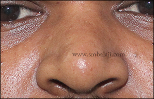
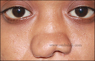
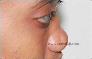
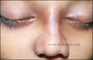
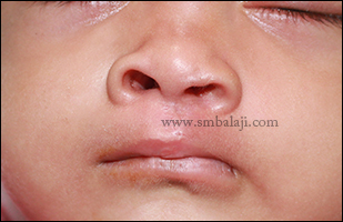
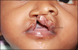
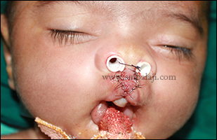
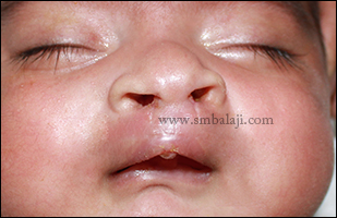
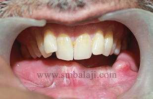
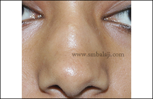
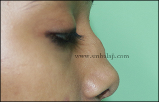
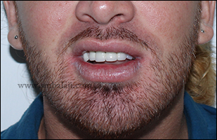
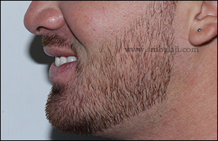
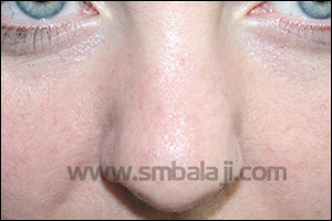
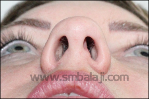
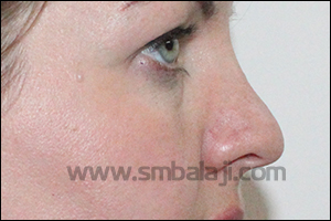
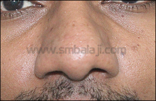
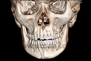
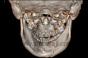
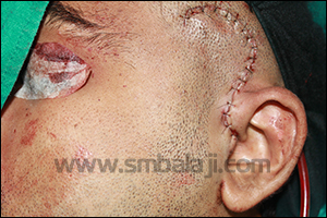
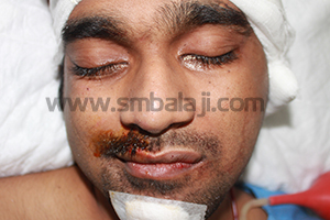
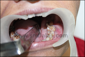
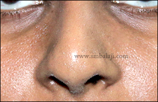
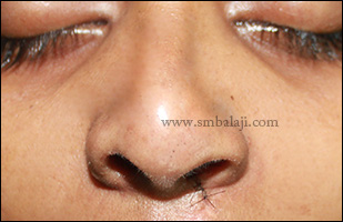
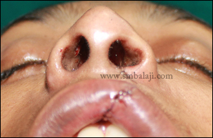
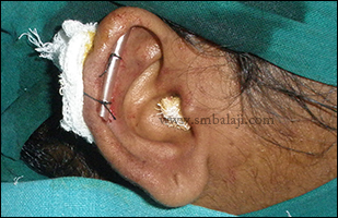
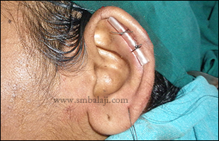
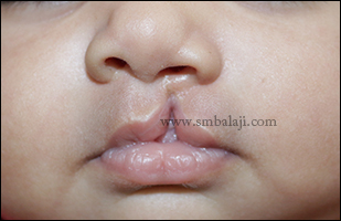
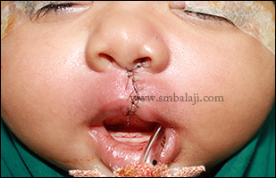
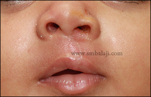
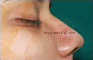
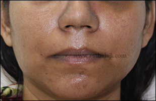
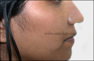
A 22-year-old man reported to our hospital seeking to correct the appearance of his nose. His nose was much flat in the middle portion and he wanted to have a sharp nose to improve his appearance.
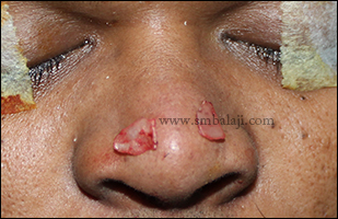
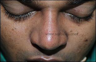
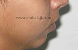
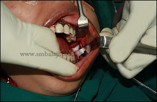
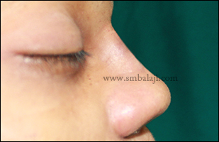
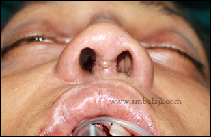
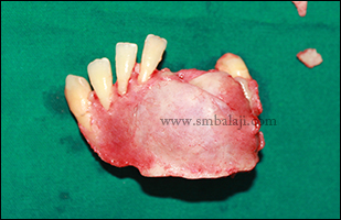
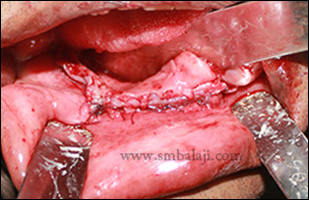
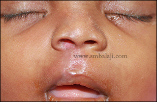
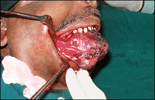
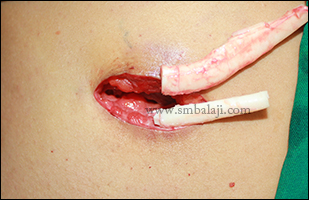
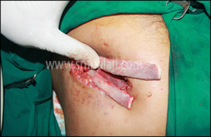
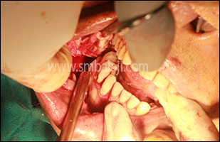
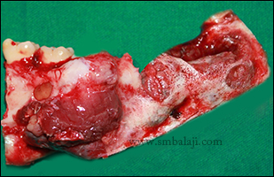
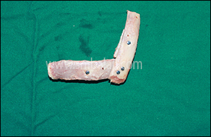
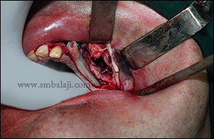
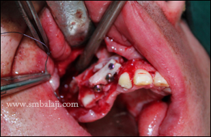
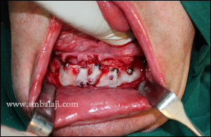
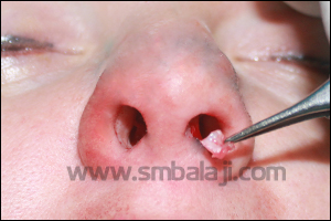
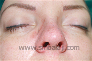
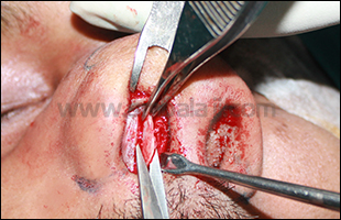
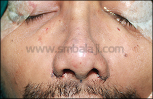
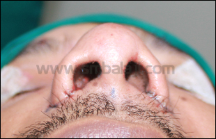
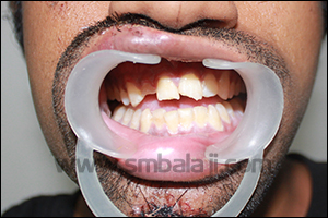
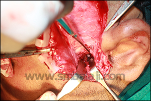
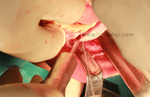
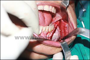
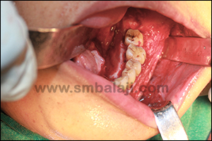
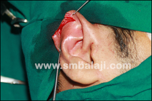
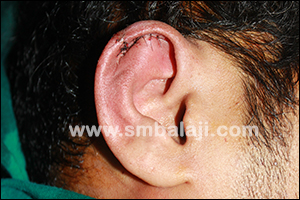
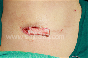
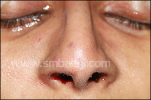
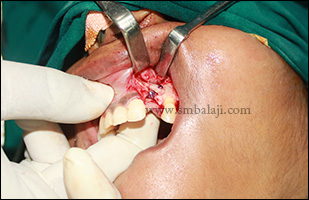
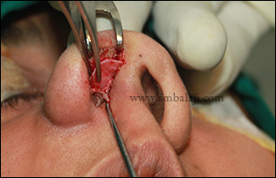
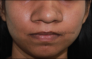
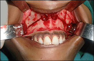
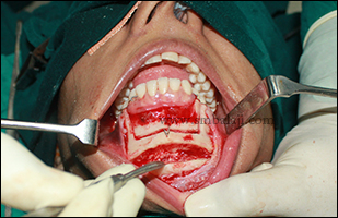
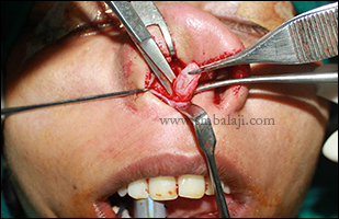
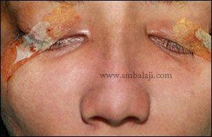
Maxillofacial surgeon Dr. S. M. Balaji expertly performed the augmentation rhinoplasty. A costochondral cartilage graft was harvested and used to reconstruct the depressed dorsum of the nose. Defined tip of the nose was achieved by removing the excessively curved portion of lower lateral nasal septal cartilage. The surgery was done from inside the nose (closed rhinoplasty) so there is no scarring. The patient is very happy to have a sharp, defined & pristine nose immediately after surgery, which improved his appearance.
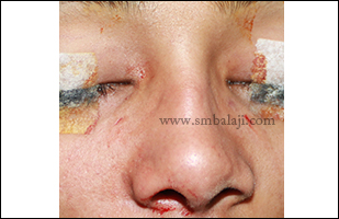
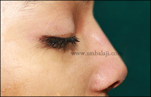
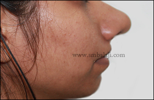
Maxillofacial surgeon Dr. S. M. Balaji expertly performed the augmentation rhinoplasty. A costochondral cartilage graft was harvested and used to reconstruct the depressed dorsum of the nose. Defined tip of the nose was achieved by removing the excessively curved portion of lower lateral nasal septal cartilage. The surgery was done from inside the nose (closed rhinoplasty) so there is no scarring. The patient is very happy to have a sharp, defined & pristine nose immediately after surgery, which improved his appearance.


 RSS Feed