Best Dental Clinic in Chennai : Best Dental Implant Center in India
FOLLOW US
  • HOME
  • Surgery of The week
  • Clinical Updates
  • Archives
  • Latest News
  • VIDEOS
  • CONTACT US

Velopharyngeal incompetence (Speech defect – hyper nasality) due to faulty pharyngoplasty corrected successfully

29/3/2014

0 Comments

 
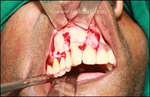
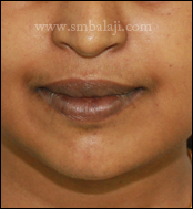
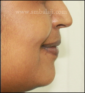
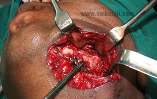
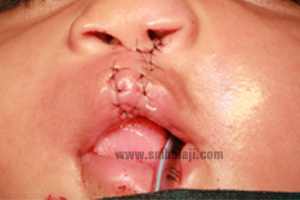
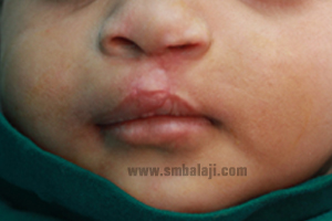
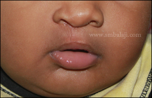
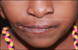
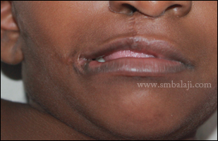
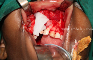
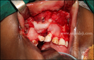
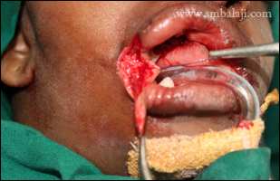
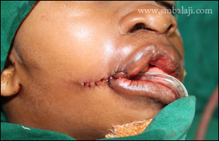
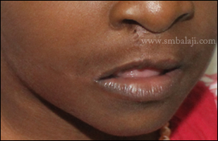
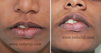
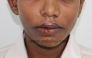
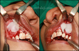
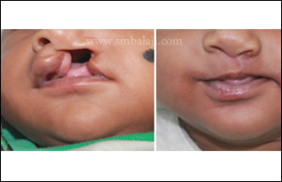
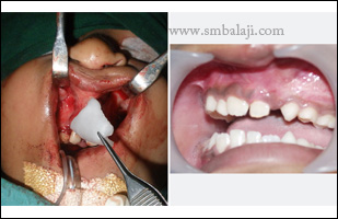
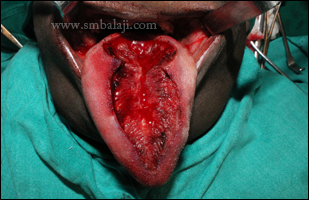
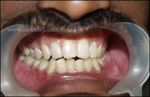
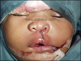
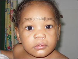
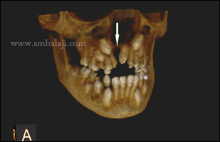
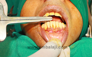
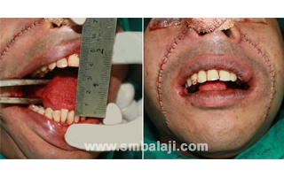
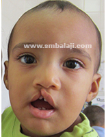
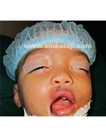
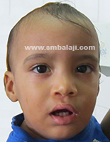
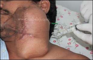
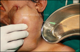
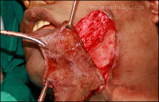
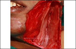
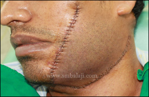
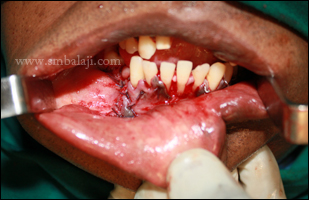
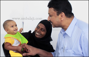
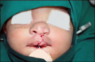
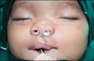
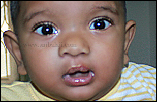
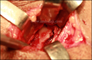
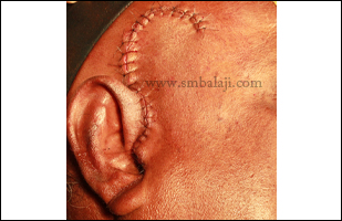
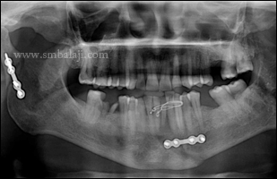
A 15 year old boy with unilateral cleft lip and palate reported to our hospital with the complaints of slurred speech. Cleft lip, palate correction and bone grating were done in their hometown during his childhood. He gave the history of flap pharyngoplasty done elsewhere 1 year back. 

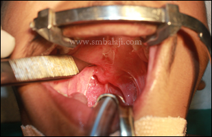
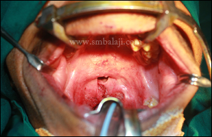
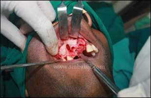
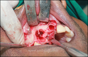
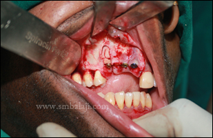
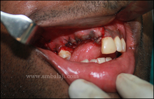
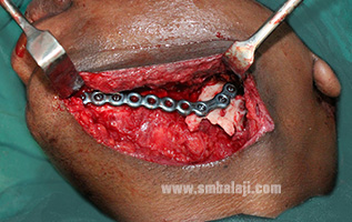
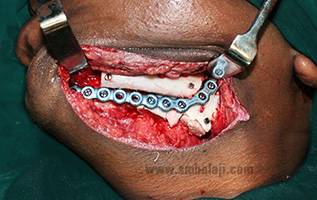
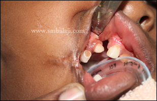
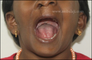
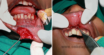
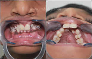
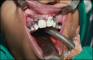
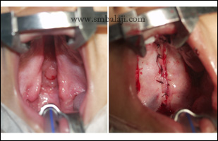
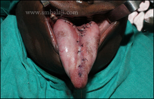
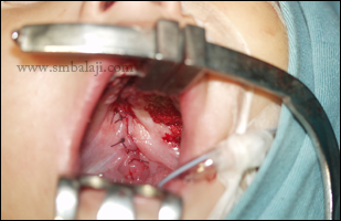
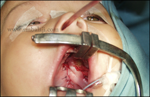
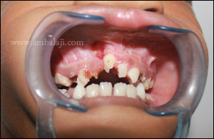
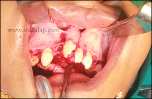
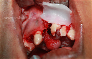
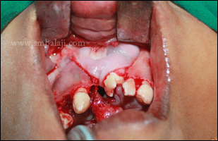
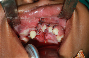
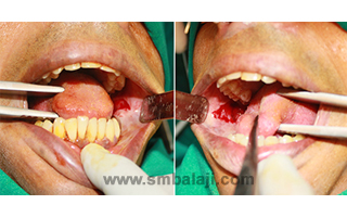
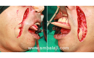
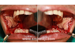
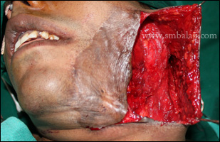
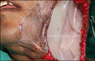
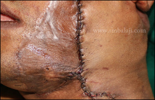
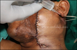
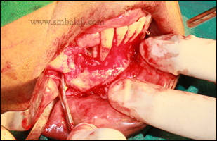
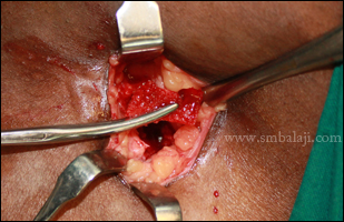
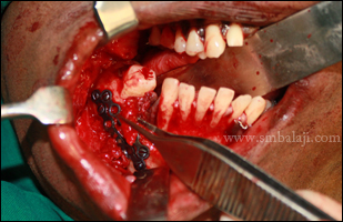
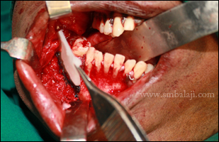
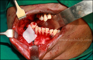
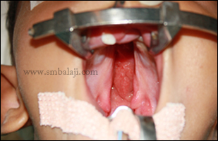
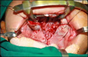
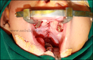
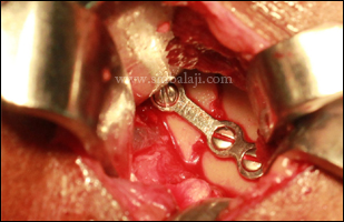
Maxillofacial surgeon Dr. S. M. Balaji skillfully diagnosed and concluded that his defective speech was due to Velopharyngeal incompetence by abnormal suturing of soft palate muscle. Thus he expertly planned to correct his speech by re positioning the pharyngeal muscles in such a way, soft palate movement will be enhanced and dynamic sphincter will be created. Thus re pharyngoplasty was performed successfully and suction test was done after the procedure which indicates positive results due to excellent soft palate movement enhancing the sphincter muscle activity. Immediate results were shown in the following video.  
Pre operative inta oral view showing improper attachment of soft palate
Post operative view showing repositioned pharyngeal muscles thus narrowing the nasal passage achieving dynamic sphincter
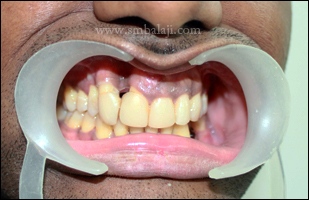
VIDEO: Intra oral video after surgery showing suction test positive thus dynamic sphincter created
0 Comments

Bone deficient region of upper jaw reinforced with bone graft & implants placed

27/3/2014

0 Comments

 
A 29-year-old man from Singapore reported to our hospital seeking expert treatment for his widely spaced upper right teeth due to which there was food impaction problem & caused an aesthetic concern. Also his upper right teeth were mobile which made it very difficult to bite & chew food. 

Digital X-ray taken showed bone loss in the region of upper right incisor, canine & first premolar that caused mobility of the teeth. Maxillofacial Surgeon Dr. S.M. Balaji performed the jaw bone reconstruction. The periodontally weakened teeth were extracted and implants placed. A symphysis bone graft was harvested from the lower jaw chin region & placed in the bone deficient region of the upper jaw to compensate the bone deficiency. The surgical site was sutured. Crown prosthesis will be given later to completely replace his missing teeth. 
Pre operative intra oral view showing spacing between the teeth due to week periodontal attachments
Pre operative digital radiograph showing severe supporting bone loss due to periodontal infection
During procedure - Periosteal flap raised showing severe bone loss
Upper anterior teeth with severe mobility was extracted
Extraction socket showing severe bone loss
Bone grafting done from the symphyseal region to enhance the bone support for implants
Implant fixed and harvested bone was placed to close the severe bone defect
Periosteal flap was sutured
0 Comments

Excellent profile enhancement by simultaneous upper & lower jaw correction

26/3/2014

0 Comments

 
A 20 year old girl reported to our hospital wanting to correct her gummy smile. Also her protruding lower jaw affected her appearance. She had low self-esteem & she wanted to enhance her looks. She had already consulted many dentists and had braces for 3 years elsewhere, but she was not satisfied with the results. Her parents too were anxious to correct her smile and enhance her facial appearance before getting her married.

Maxillofacial Surgeon Dr. S. M. Balaji expertly planned for simultaneous correction of her both upper and lower jaw. Surgical correction of gummy smile was done with Le Fort I osteotomy to reduce the maxillary excess bone. Protruding lower jaw was set back intra orally with sagittal split osteotomy. Post operatively her appearance improved greatly & instantly due to excellent profile correction. She & her parents were very happy with the immediate results & that the jaw correction surgery was done without any scars on the face. 
Pre operative frontal view showing increased exposure of teeth and incompetant lips
Lateral view showing - gummy smile with protruding lower jaw
Pre operative intra oral view showing crossbite in left side with increased vertical height of upper jaw
Post operative frontal view showing competent lips with enhanced appearance
Post operative profile view showing proportionately aligned upper and lower jaw
Post operative intra oral view showing proper bite with good overjet and overbite
0 Comments

Failed bone graft for lower jaw treated elsewhere reconstructed successfully with rhBMP-2 

26/3/2014

0 Comments

 
An 18 year old boy reported to our hospital with the complaints of severe pain and swelling in the left side of his face. Intraorally, a bone plate was exposed and huge infection with pus discharge adjacent to the exposed plate region was noticed.  He gave a history of odontogenic keratocyst (OKC) in the left side of the lower jaw which was removed elsewhere and reconstructed with bone graft and bone plate twice, 1 year back. His parents were frustrated due to repeated surgeries.

Maxillofacial Surgeon Dr. S. M. Balaji adroitly planned to remove the infected bone graft and plate and to reconstruct the lower jaw in a single surgery. He approached the surgical site extraorally through the previous scar region and removed the dislodged bone plate and sequestrum completely. Adjacent infected bone was removed with bone curette and a new NiTi bone plate was fixed to the adjacent healthy bone. To enhance the rigidity, costal graft was harvested and placed near to the bone plate with rhBMP-2 to increase the efficiency of new bone formation. Mucosal flaps were approximated in layers with very minimal scar formation. 

Post operative X-ray taken after 3 months revealed good amount of healthy bone formation maintaining the contour of the mandible using bone plate.
Pre op X ray showing failed bone grafting done elsewhere and displaced bone plate with infection
Extraoral approach through the previously operated scar region and infected bone plate removed
New NiTi bone plate fixed and stabilised well to the healthy adjacent bone
Costal graft (rib graft) harvested from his thorax region
Harvested graft fixed near the bone plate to enhance new bone formation and maintain the rigidity
Miracle protein rhBMP-2 placed to increase the efficiency of new bone formation
Mucosal flaps sutured in layers and good approximation leaving very minimal scar
Post op X ray showing good amount of healthy bone formation and maintaining the contour of the mandible with bone plate
0 Comments

Primary lip repair for unilateral cleft lip & palate

24/3/2014

0 Comments

 
A 4-month old baby girl born with unilateral cleft lip & palate was brought to our hospital by her parents seeking the best treatment for cleft defect.

Maxillofacial Surgeon Dr. S.M. Balaji performed the primary repair surgery for unilateral cleft lip using Modified Millard’s technique. Following surgery, the baby’s appearance improved greatly and she was able to feed well. The parents were very happy that their baby had hardly any post-surgical scars. Consecutively cleft palate correction surgery will be done.
Baby born with unilateral cleft lip & palate defect
Immediately after primary lip repair surgery to treat notch in upper lip
After suture removal seven days following surgery- Negligible scarring & notch in upper lip corrected
0 Comments

Successful primary unilateral cleft lip repair in a baby boy with scar less results

22/3/2014

0 Comments

 
A 3 month old baby boy born with unilateral cleft lip and palate was brought to our hospital by his parents for surgical rehabilitation of cleft deformity.

Maxillofacial Surgeon Dr. S. M. Balaji performed the primary cleft lip repair using Modified Millard’s technique. His parents were very happy that their boy’s lip defect was corrected with very minimal scar and the appearance improved greatly. The baby with his family went back to their home place and they are to return for further cleft management.

Baby boy born with unilateral cleft lip and palate
Immediate post operative view showing good approximation of cleft lip
Post operative view after 2 weeks - excellent healing with scarless results
0 Comments

Successful macrostomia correction with alveolar cleft defect repair using miracle protein rhBMP-2

21/3/2014

0 Comments

 
A girl born with unilateral cleft lip and alveolus defect along with macrostomia was brought to our hospital by her parents for surgical correction. Macrostomia is a rare type of facial cleft deformity in which the mouth is unusually wide. In this case, the girl had both alveolar cleft defect and abnormally wide mouth causing a gross distortion of her facial appearance. 

Maxillofacial Surgeon Dr. S.M. Balaji skillfully corrected the alveolar cleft defect and macrostomia in a single surgery. Alveolar cleft defect was closed with miracle protein rhBMP-2 and macrostomia was corrected by approximating the adjacent facial muscles. The defect was closed in layers and the lip was made more natural by maintaining the height and contour using the vermillion return flap technique. 

The girl’s parents felt very happy as both large mouth and cleft repair surgery was combined and post operatively scars were very less. 
Patient with abnormally large mouth (macrostomia or lateral facial cleft)
Pre operative view showing lateral facial cleft on right side of face
Alveolar cleft in the tooth bearing region of upper jaw
Miracle protein rhBMP-2 used for alveolar cleft repair
rhBMP-2 stimulates new bone formation avoiding bone graft
Surgical correction of macrostomia by approximating facial muscles
Wound closure in layers
Few weeks after surgery- defect corrected and improved appearance with hardly any scar
Post operative view showing good healing with scarless appearance
0 Comments

Residual lip defect corrected following failed treatment done elsewhere

21/3/2014

0 Comments

 
A 10-year-old boy was brought to our hospital seeking specialized correction for his lip defect. They gave a history of the boy having undergone treatment elsewhere for hemangioma of both upper & lower lip. The lesion had been removed but the lip was not reconstructed properly. The thin & disproportionate upper lip caused difficulty in pronouncing certain words and also his appearance was affected.

Maxillofacial Surgeon Dr. S.M. Balaji performed the lip reconstruction surgery. The bulky lower lip was skillfully reduced and thin upper lip was corrected by increasing the fullness from the adjacent region without any graft. Thus the newly reconstructed lip was blended naturally with very minimal scars. This surgery improved his appearance and his pronunciation better. This made his parents feel very happy. 

Pre op view showing lip defect due to improper hemangioma treatment done elsewhere
During surgery - upper lip defect was corrected and approximated and lower lip swelling was removed and adjusted to normal size
On the next day of surgery - improved appearance with very minimal scar
Post op view showing enhanced appearance with scarless lip
0 Comments

Successful union of cleft bone into a single jaw bone using miracle protein rhBMP-2

19/3/2014

0 Comments

 
An 8 year old boy with bilateral cleft lip and palate defect from Mumbai was brought to our hospital with his parents for closure of the cleft defect in the teeth bearing region of upper jaw bone and further rehabilitation of his cleft deformity. The primary cleft lip and palate repair was done elsewhere.

The premaxilla was separated from rest of the alveolus which can be clearly appreciated in 3D CBCT scan. Thus upper alveolus was separated into three different parts which will severely affect the development of teeth later.

Maxillofacial Surgeon Dr. S. M. Balaji performed the premaxillary setback procedure with rhBMP-2 which helps in uniting the three parts into a single jaw bone with new bone formation. This surgery helped for proper growth and development as well as alignment of his teeth.
Pre operative intraoral view showing protruding premaxilla
3D CBCT scan showing detached and forwardly placed premaxilla from rest of the alveolus
Intra oral view showing severe anterior deep bite due to lowered position of premaxilla and palatally placed lateral incisors due to lack of bone
During surgery rhBMP-2 placed in the cleft defect region
Immediate post operative view showing accurate approximation of premaxilla
0 Comments

Complete rehabilitation for cleft child from 3 months to 16 years of age Successive surgeries with 16 years follow-up

18/3/2014

0 Comments

 
Sixteen years ago, a 4-month-old baby girl born with unilateral cleft lip & palate was brought to our hospital by her parents for the complete rehabilitation of her cleft deformity. Maxillofacial Surgeon Dr. S.M. Balaji performed the successive surgeries over the years.

Primary cleft lip repair surgery was done when the baby was 4 months old and palate repair surgery was done at 8 months of age. When the baby was 6 years old, rhBMP-2 surgery was done to treat the alveolar cleft defect. Miracle protein rhBMP-2 was placed in the cleft of the teeth bearing region of the upper jaw to completely heal the bony defect avoiding bone graft.

Now the girl reported to our hospital seeking to enhance her facial profile. She had a hypoplastic maxilla (retruded upper jaw) and she was not happy with her appearance. Dr. Balaji performed the intraoral maxillary distraction osteogenesis whereby the upper jaw was advanced and aligned properly with the lower jaw. This was done completely inside the mouth so there were no scars. Using distraction & orthodontics, her appearance & facial profile was greatly enhanced.  
0 Comments

Surgical trimming of large tongue

17/3/2014

0 Comments

 
A 19-year-old boy from Trichy was brought to our hospital by his parents for the surgical correction of his abnormally large tongue due to which he had difficulty in speaking and closing his mouth. 

This condition is called Macroglossia. On examination, the boy had no associated symptoms or any other systemic illness. The surgery was carefully planned & meticulously executed by Maxillofacial Surgeon Dr. S.M. Balaji. The large tongue was surgically trimmed with utmost care taken not to damage the huge blood vessels inside the tongue. A modified resection design incorporating the Pichler's central reduction, modified key-hole, and Kole's approach were incorporated to form a rough clover shaped design. Following this technique, both the length and width of the tongue were reduced more precisely. By choosing this design, the sensitive nerves and the tip of the tongue were preserved so as not to compromise taste perception. The boy is now able to use the new normal sized tongue to speak, swallow and taste. 

Pre operative view showing large tongue leading to inability to close the mouth
Surgical reduction of both length and width of the tongue using modified key hole technique
Immediate post operative view showing reduction of tongue to a normal size
Post operative view showing normal size tongue and able to close the mouth efficiently
0 Comments

Bilateral condylar head fracture management

15/3/2014

0 Comments

 
A 28-year-old software engineer was brought to our hospital after he sustained injuries to his face in a road traffic accident. After immediate first aid, he complained of pain in the jaws and inability to bite properly. 

A 3D CT scan taken showed a fracture in both the right & left mandibular condylar heads and both fractured segments were displaced inwards. This caused a derangement in the patient’s occlusion or contact between upper and lower teeth. Maxillofacial Surgeon Dr. S.M. Balaji performed the surgical fracture reduction. The fracture site was surgically exposed, the patient’s occlusion was accurately restored, the fractured segments were stabilized and fixed in their normal position with bone plates and anterior open bite was corrected. 

0 Comments

Isolated cleft palate repair

15/3/2014

0 Comments

 
A baby boy born with isolated cleft palate defect was referred to our hospital for surgical correction of the cleft defect.

Maxillofacial Surgeon Dr. S.M. Balaji performed the cleft palate repair using Palatal pushback technique. The palatal flap was raised on both right & left side. The palate muscles were reattached in their normal position & a two layer closure was done to completely reconstruct the floor of the nose and roof of the mouth. The suction test was positive which guarantees good speech.

Isolated cleft palate defect
Immediately after palate repair surgery
Suction test was performed
0 Comments

Primary lip repair for African baby girl

13/3/2014

0 Comments

 
An 8-month-old baby girl from Africa, born with unilateral cleft lip & palate, was brought to our hospital by her parents for the surgical correction of her lip defect.

Maxillofacial Surgeon Dr. S.M. Balaji performed the primary lip repair surgery using the Modified Millard’s technique. The baby’s parents were very happy that their baby’s lip defect was corrected with hardly any scars and the child’s appearance greatly improved.  The baby and her family went back to her homeland and they are to return later for further cleft management. 

Baby girl born with unilateral cleft lip and palate
Immediately after primary cleft lip repair surgery
Few weeks after surgery- negligible scar on lip and improved appearance
0 Comments

Alveolar cleft repair using rhBMP-2 avoiding bone graft

12/3/2014

0 Comments

 
An 8-year-old boy was brought to our hospital by his parents for surgical correction of his cleft defect. His cleft lip and cleft palate defect was operated elsewhere, in his childhood. The surgery results were adverse as the entire pre-maxilla was removed.

Maxillofacial Surgeon Dr. S.M. Balaji performed the alveolar cleft repair using rhBMP-2 avoiding bone grafting. Miracle protein rhBMP-2 is a bioengineered version of a protein that occurs in the body. When placed in the cleft defect, it stimulates the body’s stem cells to form new bone and thereby healing the bony defect in the teeth bearing region of the upper jaw. By using rhBMP-2, bone graft surgery from the hip or rib is avoided, thereby avoiding an additional surgery & unsightly scars. 
Alveolar cleft defect
3D CBCT scan showing alveolar cleft defect in right side upper jaw
Alveolar cleft region surgically exposed
rhBMP-2 placed in alveolar cleft avoiding bone graft
rhBMP-2 stimulates new bone formation ultimately healing the bony defect
Immediately after surgery-surgical site sutured
0 Comments

Oral Submucous Fibrosis

11/3/2014

0 Comments

 
A 32-year-old man came to our hospital seeking treatment for his extreme inability to open his mouth normally. Preoperatively, his mouth opening was just 11 mm. He gave a history of cigarette smoking and pan chewing for 2 decades. He was diagnosed with Oral submucous fibrosis, a condition characterized by thickening of the mucosal tissues and progressive rigidity of the jaws making it exceedingly difficult to open the mouth. Previously he underwent laser surgery elsewhere, 3 times, for this ailment but it did not work. 

In the surgical treatment procedure, Maxillofacial Surgeon Dr. S.M. Balaji placed transverse incisions on the cheek on both right & left side to release the fibrous bands and to immediately improve the mouth opening. An inferiorly based Nasolabial island flap based on the facial artery was raised and transposed intraorally through a small trans-buccal tunnel. This was sutured with the buccal mucosal defect. Extraorally, the defect was closed in layers. Immediately following surgery, a normal mouth opening of 35 mm was achieved.

Severely limited mouth opening before surgery
Fibrous bands released on left & right cheek to improve mouth opening
Nasolabial island flap based on facial artery raised & transposed intraorally on both sides
Flaps sutured with the buccal mucosal defect on right & left side
Following surgery, normal mouth opening achieved with minimal scarring
0 Comments

Successful primary cleft lip repair surgery

10/3/2014

0 Comments

 
Nine-month-old Mohammed Altaf, from UAE, was born with incomplete cleft lip. His father & mother brought him to our hospital for specialized cleft care.

Maxillofacial Surgeon Dr. S.M. Balaji performed the primary cleft lip repair surgery using Modified Millard’s Technique. Following surgery, there was very negligible scar on the upper lip and the boy’s appearance improved greatly. This made the boy’s parents feel very happy with the surgery success. 

Boy from UAE born with incomplete cleft lip
Immediately after primary cleft lip repair
Few weeks after surgery - negligible scar
Dr. S.M. Balaji with the boy
0 Comments

Augmentation rhinoplasty for enhanced appearance

8/3/2014

0 Comments

 
A 35-year-old woman reported to our hospital seeking to enhance the appearance of her nose. Her nose was flattened and ill-defined making her very self-conscious about her appearance. She wanted to improve her facial profile.

Maxillofacial Surgeon Dr. S.M. Balaji performed a nose correction procedure called Augmentation Rhinoplasty. The nasal bridge was raised with a cartilage graft (costal cartilage). The surgery was done within the nose so there are no scars on the face. The woman’s nose was made sharp & pristine that greatly enhanced her appearance & profile. She was very happy to have a renewed confidence with a new, improved look.
Flattened, ill-defined nose
Profile view before surgery
Surgical planning with costal cartilage
Nose made sharp & pristine without scars following augmentation rhinoplasty
Enhanced appearance in profile view following nose correction
0 Comments

Successful reconstruction of facial burn injury using Tissue Expansion technique

7/3/2014

0 Comments

 
A 28-year-old electrician came to our hospital seeking expert treatment for his face burn. He had suffered a severe burn injury to the left side of his face, in an accident at his workplace. Elsewhere, a split-thickness skin graft surgery from his thigh was done but the patient was very unhappy with the graft shrinkage and color mismatch.

Maxillofacial Surgery Dr. S.M. Balaji performed the facial reconstruction surgery using the novel “Tissue expansion” technique wherein a device called tissue expander was placed beneath the normal skin on the left cheek. Saline solution was infused into the device regularly. The device gradually expanded, stretching the overlying skin. New, healthy skin was grown at the site locally, of the same color & texture. This skin was used to reconstruct the scarred portion of the left cheek, chin & neck. Since the tissue was grown locally, there was no color mismatch. The wound healed well and his appearance improved drastically
Video showing saline being drained out from the expanded device before the device is removed
0 Comments

Lower jaw fracture successfully treated after previous failed procedures done elsewhere

6/3/2014

0 Comments

 
CT scan showing non-healing results of previous surgeries done elsewhere for right side lower jaw fracture
Fracture site exposed showing non-union of segments
Fracture fixed with bone plates and screws
Cortico-cancellous bone graft harvested from right hip region
Bone graft packed into the fracture defect
Bone ends covered with collagen sponge soaked in rhBMP-2
rhBMP-2 will stimulate new bone formation at the bone ends ensuring complete healing
Immediately after surgery- Wound site sutured
A 35-year-old man reported to our hospital seeking expert treatment for his lower jaw (mandible) fracture. He gave a history of having sustained the injury in a road traffic accident. He had sought various treatments elsewhere but the multiple surgeries were futile with non-healing results. 

A CT scan showed complete non-union of the fractured segments. Maxillofacial Surgeon Prof. S.M. Balaji performed the surgery for jaw fracture fixation. The fracture site was accessed and fibrous tissue as a result of non-healing of the fractured fragments was removed. The bone ends were freshened, proper occlusion was established, and the fractured fragments of the lower jaw were fixed with a 6-hole & 4-hole bone plate with 2 mm screws. A cortico-cancellous bone graft from the right hip region was harvested and packed into the fracture defect. The graft and bone ends were covered with a collagen sponge soaked in rhBMP-2. rhBMP-2 is a miracle protein that stimulates the body’s own cells to form new bone thereby ensuring complete healing of the bone defect. Following this, the wound was sutured.

0 Comments

Cleft palate repair

5/3/2014

0 Comments

 
A baby boy born with cleft lip & palate was previously operated by Maxillofacial Surgeon Dr. S.M. Balaji for primary cleft lip repair. The baby now 9 months old, was brought to our hospital by his parents for surgical correction of the cleft palate.

Dr. Balaji performed the cleft palate repair using Palatal pushback technique. Incision was made in the mid-vomerine region. The palatal flap was raised on both right & left side. The Levator palatine muscles were detached from their abnormally attached positions and reattached into normal position like a hammock. A two layer closure was done. The nasal floor was closed with the vomerine flap making a reverse knot. Oral layer was sutured by vertical mattress sutures. The vertical mattress sutures produce a ridge of thick mucoperiosteum as the flaps are approximated to each other in the midline. The suction test was positive which guarantees good speech.
Cleft palate defect
Palatal flap raised on both sides
Levator muscle reattached into normal position like a hammock
Complete two layer closure done for palate repair
0 Comments

Primary cleft lip repair for Sri Lankan baby

4/3/2014

0 Comments

 
A 6-month-old Sri Lankan baby boy, born with unilateral cleft lip and palate, was referred to our hospital for surgical correction of his cleft defect.

Maxillofacial Surgeon Dr. S.M. Balaji performed the primary cleft lip repair surgery. Following lip correction, the baby is able to feed well and his facial appearance has improved greatly. In the near future, correction of his palate defect will be done for complete rehabilitation. 

Unilateral Cleft lip & palate
At consultation
During primary cleft lip repair surgery
Immediately after surgery
After suture removal on 7th post-operative day
Second week post-operative appearance
0 Comments

Successful correction of abnormal tear flow from eye (epiphora) by Dacryocystorhinostomy with medial canthopexy 

4/3/2014

0 Comments

 
A 34-year-old lady reported to our hospital with a complaint of continuous tears running down her face from her left eye. She gave a history of having met with a car accident.  Due to the traumatic injury, it was identified that the medial canthal ligament of her left eye had detached and come down. A defect in her nasolacrimal duct caused epiphora or abnormal tear flow. The normal flow of tears from the eye to the nose was damaged.

The aim of the surgery was to correct the defect in the nasolacrimal duct with DCR or Dacryocystorhinostomy & reposition the medial canthal ligament. The ostium was opened with rongeurs, bone adjacent to nasolacrimal sac was removed. A new direct communication was created between the nasolacrimal sac & the nasal mucosa by placing a silicone tube to enable the tears to drain into the nose. The medial canthal ligament was repositioned using Y-plate canthopexy. 
Displaced medial canthus due to traumatic injury
Dacryocystorhinostomy surgery for correction of abnormal tear flow
Ostium opened and direct communication created between nasolacrimal sac & nasal mucosa using silicone tube
X-ray taken after surgery showing Y plate used to reposition medial canthal ligament
Medial canthal ligament repositioned and epiphora corrected following DCR surgery
0 Comments

Fracture of lower jaw successfully treated

1/3/2014

0 Comments

 
Preoperative radiograph showing mandible fracture and condyle fracture
Preauricular endaural approach for open reduction of condyle fracture
Fracture site approached
Fracture fixation using bone plate
Wound closure immediately after surgery
Postoperative radiograph showing good fracture fixation
A middle-aged man sustained severe injuries to his lower jaw in a road traffic accident. He was rushed to our hospital for emergency care & treatment.

A digital radiograph showed a fracture in his lower jaw in the front teeth region & a fracture of the right jaw joint. In the open reduction surgery performed by Maxillofacial Surgeon Dr. S.M. Balaji, the right jaw joint or temporomandibular joint was surgically exposed through a preauricular endaural approach. The condyle fracture segments were aligned in their normal position & fixed with bone plate. The fracture in the middle of the lower jaw was also fixed with bone plate. Subsequently, the fractures healed well.
0 Comments

    Categories

    All
    Abbe Flap Technique
    Advanced Transport Distraction Osteogenesis
    Advancement Genioplasty
    Alveolar Cleft Repair
    Alveolar Distraction
    Ameloblastoma
    Augmentation Genioplasty
    Augmentation Rhinoplasty
    Bat Ear Correction
    Bilateral Cleft Lip Palate
    Blepharoplasty
    Bone Defect Reconstruction
    Bone Grafting
    Bone Morphogenetic Protein(BMP)
    Broad Flattened Nose Correction
    Bulky Nose Correction
    Chin Correction
    Chin Correction Surgery
    Cleft Lip & Palate Surgery
    Cleft Nose
    Cleft Palate Repair
    Cleft Rhinoplasty
    Complete Facial Rehabilitation
    Complex Facial Fractures
    Condylar Fracture Surgery
    Correction Of Hypoplastic Ramus
    Corrective Jaw Surgery
    Cosmetic Cleft Nose Correction
    Cosmetic Nose Correction
    Cosmetic Rhinoplasty
    Cosmetic Surgery
    Craniofacial Deformities
    Craniofacial Reconstruction
    Craniofacial Surgery
    Cyst Removal
    Dental Implant
    Dentigerous Cyst
    Deviated Nose Correction
    Diplopia
    Distraction Osteogenesis
    Ear Correction
    Ear Defect
    Ear Reconstructive Surgery
    Epiphora
    Extruding Infected MEDPOR
    Eyebrow Correction
    Face Enhancement
    Face Reconstruction
    Facial Asymmetry Correction
    Facial Bone Fracture
    Facial Burn Injury
    Facial Palsy Surgery
    Fibrous Dysplasia
    Flat Nose Correction
    Forehead Bone Fracture
    Forehead Deformity
    Forehead Flap
    Frontal Bossing Correction
    Gummy Smile
    Hemifacial Microsomia
    Hypertelorism Surgery
    Implant
    Incomplete Cleft Lip
    Isolated Cleft Palate Repair
    Jaw Correction Surgery
    Jaw Cyst
    Jaw Deformity
    Jaw Fracture Surgery
    Jaw Joint Ankylosis
    Jaw Reconstructive Surgery
    Jaw Surgery
    Lip Correction
    Lip Defect Correction
    Lip Revision
    Locked Jaw
    Lower Jaw Fracture
    Low Lying Upper Eyelid Correction
    Macrostomia Correction
    Mandible Correction
    Mandible Reconstruction
    Maxillofacial Surgery India
    Maxillomandibular Distraction
    Maxilofacial Surgery
    Micrognathic Mandible Correction
    Microtia
    Nasal Glial Heterotopia
    Naso-orbito-ethmoidal Fracture
    Nose And Lip Correction
    Nose Asymmetry Correction
    Nose Correction
    Nose Job
    Nose Reconstructive Surgery
    Oral Submucous Fibrosis
    Oral Tumor Removal
    Orbital Correction
    Orehead Defect
    Orthognathic Surgery
    Parrot-beak Nose
    Parry Romberg Syndrome
    Pharyngoplasty
    Plastic Surgery
    Primary Cleft Lip Repair
    Profile EnhancemeP
    Reduction Glossectomy
    Reduction Rhinoplasty
    Removal Of Capillary Hemangioma
    RhBMP 2
    RhBMP-2
    Rhinoplasty
    Road Accident Trauma
    Road Accident - Trauma
    Scar Revision
    Secondary Cleft Palate
    Sinus Lift
    Smile Makeover
    Speech Correction
    Square-face Correction
    Surgery Without Scar
    Tessier Nose Defect
    Tissue Expansion
    TMJ Ankylosis
    Tongue Correction
    Tongue Surgery
    Trauma
    Trauma Treatment
    Triangular Shaped Forehead
    Tumor Resection
    Unilateral Cleft Lip
    Unilateral Cleft Lip And Palate
    Unilateral Cleft Lip Repair
    Velopharyngeal Incompetence

    Archives

    April 2021
    March 2021
    January 2021
    December 2020
    April 2020
    March 2019
    February 2019
    January 2018
    November 2017
    October 2017
    September 2017
    June 2017
    May 2017
    April 2017
    March 2017
    January 2017
    December 2016
    October 2016
    September 2016
    August 2016
    July 2016
    June 2016
    May 2016
    April 2016
    March 2016
    February 2016
    January 2016
    December 2015
    October 2015
    September 2015
    August 2015
    July 2015
    June 2015
    May 2015
    April 2015
    March 2015
    February 2015
    January 2015
    December 2014
    November 2014
    October 2014
    September 2014
    August 2014
    July 2014
    June 2014
    May 2014
    April 2014
    March 2014
    February 2014
    January 2014
    December 2013
    November 2013

    RSS Feed

Copyright © 2020 Balaji Dental & Craniofacial Hospital | All rights reserved | Powered by BALAJI DENTAL HOSPITAL