Best Dental Clinic in Chennai : Best Dental Implant Center in India
FOLLOW US
  • HOME
  • Surgery of The week
  • Clinical Updates
  • Archives
  • Latest News
  • VIDEOS
  • CONTACT US

Successful treatment of lower jaw fractures sustained in a vehicular accident

28/11/2014

0 Comments

 
Preoperative 3D CT scan image showing left mandibular angle fracture
OPG of the patient showing the two mandibular fracture lines
Completely deranged occlusion due to fracture causing inability to bite, chew, swallow, speak
Fracture segments stabilized with bone plate and screws on the right mandible
Left side fracture stabilized and plated
Proper occlusion and bite achieved with successful treatment
Postoperative radiograph showing successful fixation and stabilization of fractures immediately after surgery
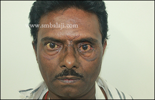
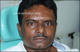
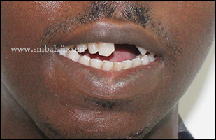
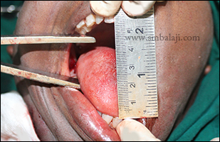
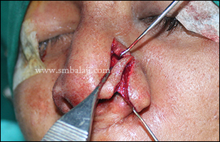
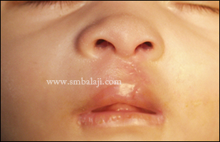
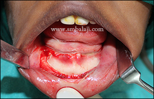
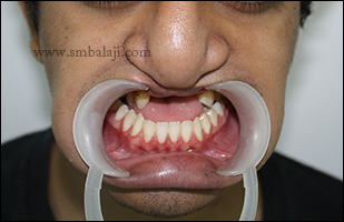
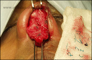
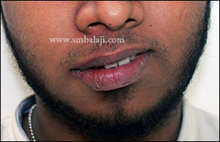
A young man had met with a motor vehicular accident a few days in which he had sustained injuries to his lower jaw. He had severe jaw pain, his teeth occlusion was completely deranged and he had great difficulty in biting, chewing, swallowing and speaking.

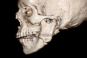
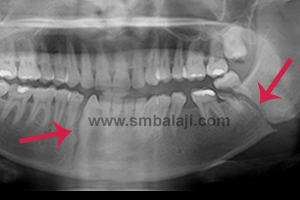
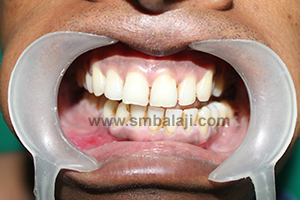
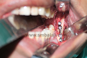
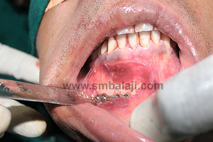
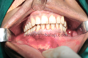
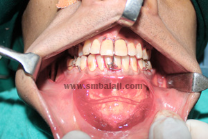
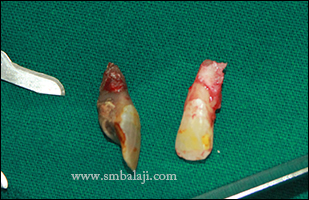
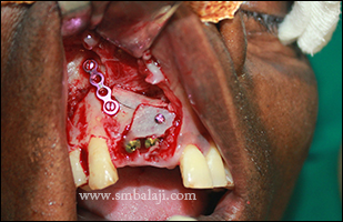
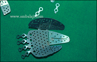
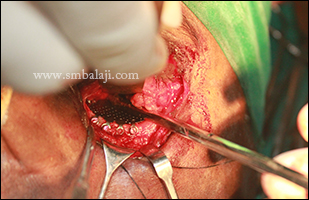
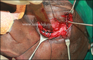
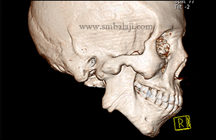
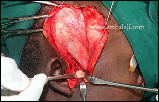
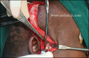
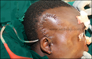
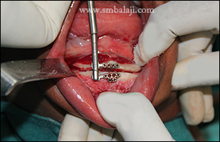
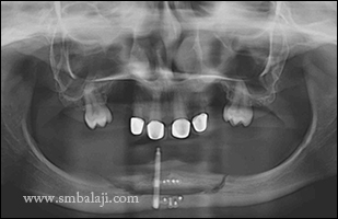
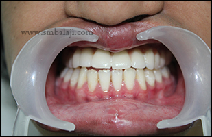
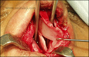
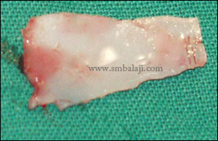
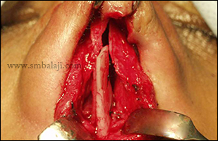
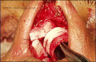
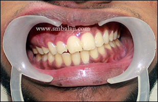
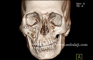
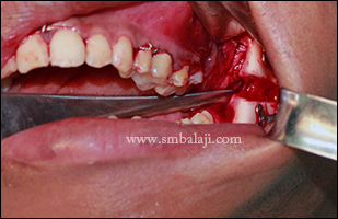
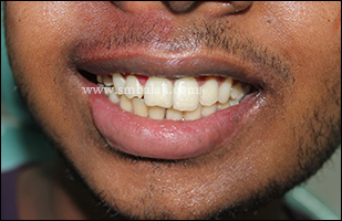
A preoperative 3D CT scan revealed fracture in the right mandibular parasymphysis region and left angle of mandible. Maxillofacial Surgeon Dr. S.M. Balaji successfully fixed and stabilized the fractured segments using bone plates and screws. The fractured tooth in the line of fracture was extracted. Accurate occlusion and bite was achieved. Postsurgery radiograph showed optimum fracture fixation. 
0 Comments

Combined lower jaw and chin correction for enhanced appearance

27/11/2014

0 Comments

 
A young man came to our hospital seeking corrective jaw surgery to enhance his appearance. His lower jaw was excessively protruding causing an incorrect teeth occlusion that caused difficulties in biting and chewing. Additionally his chin bone was deviated away from the midline giving his face an asymmetric, crooked appearance.

Maxillofacial surgeon Dr. S.M. Balaji planned to correct the jaw defect with a combined bilateral Obwegeser’s sagittal split osteotomy and genioplasty. The mandible was set back to correct the alignment and bite using bilateral Obwegeser’s sagittal split osteotomy. With genioplasty, the deviated chin bone was corrected. The procedure was done from inside the mouth so there were no scars. Immediately after this corrective jaw surgery, the man’s appearance was greatly improved and he is very happy with the enhanced aesthetics.
Crooked chin giving face an asymmetric appearance
Incorrect bite due to protruding lower jaw and deviated chin bone
Combined bilateral Obwegeser sagittal split osteotomy (lower jaw correction) and genioplasty (chin correction)
Asymmetry and deviated chin corrected improving appearance immediately after surgery without any scars
0 Comments

Double vision (Diplopia) correction of orbital blow out – eye bone fracture

24/11/2014

0 Comments

 
A 42 year old man from West Bengal reported to our hospital for treatment of fractures of his face bones that he had sustained in a vehicular accident. His 3D CT scan showed right Le Fort I fracture & nasal bone fracture. Also there was left orbital blow out fracture and fracture of the lateral orbital wall on the left side. The patient had enopthalmus (sunken eyes) and diplopia (double vision) in the left eye.

Maxillofacial Surgeon Dr. S. M. Balaji successfully performed the facial fracture rehabilitation. Under general anesthesia, iliac graft was obtained from the medial cortex using trap door technique. Upper central and lateral incisors were extracted; implants were placed in the freshly extracted sockets.  To compensate for the bony defect, iliac bone was placed and fixed using screws. To address the orbital floor fracture, transconjuctival incision was placed with lateral canthotomy. The fracture along the frontozygomatic suture and lateral orbital wall was stabilized using bone plates and screws, the herniated orbital contents were then retracted. Titanium mesh contoured, adapted over the floor and fixed using screws. Iliac graft placed over the mesh.  The nasal bone was elevated and a nasal pack placed. On the 3rd post-operative day the patient demonstrated full range of ocular movements with regression of double vision. 
Preoperative facial view showing enolphthalmus and asymmetrical appearance and sunken left eye leading to diplopia
Preoperative 3DCT scan view showing fractured orbital floor and lateral wall with depressed orbital floor giving altered levels of orbit
By following trap door technique cortical bone was obtained from iliac crest
Infected teeth from upper front region of the jaw was extracted
Nobel implant placed in the missing teeth region and bone graft was fixed and implant was completely covered and stabilized
Titanium mesh was used to reconstruct the orbital floor
Titanium mesh was fixed in the floor of the orbit and sunken orbital content was raised from the fractured bone segments
Iliac bone graft was used to raise the orbital floor and fractued lateral orbital wall also fixed with mini boneplate
Postoperative view after 2 weeks showing corrected orbit level and lateral orbital wall positioned giving enhanced appearance
0 Comments

Successful correction of TMJ ankylosis with Gap arthroplasty

21/11/2014

0 Comments

 
This was a case of unilateral TMJ ankylosis in a 24 year old boy from Nigeria. He was not able to open his mouth even for taking food for past 18 years due to an accident. Following the injury, his upper front teeth were lost and he was taking food only through this small gap. 3DCT scan shows TMJ bony ankylosis on the right side and elongated coronoid processes.

Maxillofacial Surgeon Dr. S. M. Balaji planned to open his mouth by following gap arthroplasty with temporalis fascia interpositioning and contralateral coronoidectomy. Under GA, Al-Kayat-Bramley’s incision (Preauricular incision) was made on the right side; a superior limiting cut was made along the demarcation between the rim of the glenoid fossa and the ankylosed head. The ankylotic mass was removed. After ressecting the callous, the gap was further widened by removing adequate bone.

Intraoral coronoidectomy on the contralateral side i.e. the left side was done to facilitate good mouth opening. On the right side, the temporalis muscle was rotated and folded inwards into the joint cavity and taken between the medial surface of the ramus taking good care of the adjacent mandibular nerve, facial and maxillary arteries. Then the muscle end was sutured to the medial side of the mandible to provide a submandibular anchorage. A suction drain is placed for 24-36 hrs and the wound was closed in layers. Results are immediate and mouth opening of inter-incisor width up to 55mm was achieved.
Preoperative view showing negligible mouth opening with missing upper left central and lateral incisors
3DCT scan showing bony fusion of right side condyle with the base of the skull
Preauricular incision in the right side visualizing the ankylosed condyle with that of the glenoid fossa
Exactly in the demarcation between condylar head and glenoid fossa bone cut was made
Temporalis muscle with fascia was detached and interpositioned between condylar head and glenoid fossa and sutured medially in the submandibular region
Immediately after layer closure of the preauricular incision drain out was attached for 24-48 hours to prevent hematoma formation
Immediately after contralateral coronoidectomy mouth opening was increased upto 55mm
0 Comments

Extruding infected MEDPOR from treatment done elsewhere removed successfully from the eye socket

19/11/2014

0 Comments

 
An African boy with Parry Romberg syndrome previously treated elsewhere reported to our hospital with a complaint of excruciating pain near his right eye. He had undergone reconstruction of his orbit (eye socket bone) elsewhere using MEDPOR few months back which had now become infected and was extruding through the skin close to his right eye. There was pus discharge and caused him severe pain.

Maxillofacial Surgeon Dr. S.M. Balaji successfully removed the extruding, infected MEDPORE from the orbit. The surgical site was sutured and he was put on medication to treat the infection. By timely removal of the failed MEDPOR and controlling the infection, the serious risk of his vision getting affected has been averted. 
Extruding infected MEDPOR in orbit in African boy
Infected MEDPOR from the treatment done elsewhere removed
All implants removed without damage to surrounding vital structures
The infected MEDPORE removed successfully
Surgical site sutured
0 Comments

Nose asymmetry corrected with rhinoplasty

15/11/2014

0 Comments

 
A man with Tessier nose defect came to our hospital wanting to surgically change the shape of his nose to improve his appearance. His nostrils were asymmetrical with the left side being at a higher level than the right. The nasal septum was deviated.

Maxillofacial surgeon Dr. S.M. Balaji successfully performed the rhinoplasty to correct the alar deficiency. Deviated septum was corrected. Footplate of the medial crus on the left side was removed. Using Denonvillier’s Z-plasty technique lower lateral cartilage was brought down to reduce the columellar show on the left side. Both nostrils were made symmetrical improving the appearance of the nose and enhancing the facial profile. 

Preoperative view showing Tessier's cleft with defective ala of the nose in the left side
Preoperative view showing reduced size of the nostril in the left side with uneven alar base
Footplate of medial crus removal in the left side which reduces the columella show
Z plasty technique with Denonvillier's incision the alar level was adjusted and ala of the nose in the left side was pushed down
Immediately after suturing shows corrected alar base with symmetrical appearance of the nose
Postoperative view showing symmetrical nostrils and corrected shape of the ala of the nose
0 Comments

Unilateral cleft lip repair

14/11/2014

0 Comments

 
A baby girl born with unilateral cleft lip and palate was brought to our hospital by her parents for management of cleft deformity. Maxillofacial Surgeon Dr. S.M. Balaji performed the primary repair of the unilateral cleft lip defect using modified Millard’s technique. Suture removal was done seven days following surgery. The lip defect was successfully corrected with hardly any scars greatly improving the baby’s appearance. Further cleft surgeries will be done at appropriate stages. 
3 months old baby girl with unilateral cleft lip defect
Immediately after primary cleft lip repair
Immediate postoperative view after suture removal following seven days after surgery
2 months after primary cleft lip repair showing enhanced appearance with hardly any scars on the upper lip
0 Comments

Alveolar distraction to increase height of chin bone to enhance appearance

13/11/2014

0 Comments

 
Ectodermal dysplasia is a rare genetic condition characterized by abnormal development of skin, hair, nails, teeth and sweat glands. A young girl suffering from this condition came to our hospital seeking a fixed replacement of her missing teeth and to correct the shape of her chin which gave her an aged appearance.

A 3D CT scan was taken to gauge the amount of residual bone. Due to the multiple missing teeth over a long period of time, the lower jaw bone was severely resorbed and the vertical height was insufficient to place implants. Maxillofacial Surgeon Dr. S.M. Balaji planned for a surgical reconstruction using alveolar distraction osteogenesis technique. The anterior region of the lower jaw bone was cut and alveolar distractor device was fixed. When the screw attached to the device is gradually turned 0.6 mm daily, the cut segments of bone move apart and new bone is formed in the resultant gap ultimately increasing the bone height which enhances her appearance. 
Preoperative 3DCT view showing reduced height of the alveolar bone in the front region of the lower jaw due to congenitally missing teeth
Preoperative digital OPG showing reduced height of alveolar bone in the lower front region of the mandible
Intraoral view showing reduced height of the alveolar process with illdefined vestibular sulcus in lower anterior region
During procedure mucoperiosteal flap raised to expose the alveolar process of the mandibular anterior region
Bone cut was placed superior to the mental foramen and segmental alveolar distractor was fixed to it
Trial activation was done and movement of the alveolar segment was checked
Mucoperiosteal flap was approximated with sutures
1 week postoperative digital OPG showing moved alveolar segment to increase the height of the chin region
0 Comments

Rhinoplasty for correction of cleft nose defect

11/11/2014

0 Comments

 
A girl with unilateral cleft lip and palate came to our hospital seeking to correct her nose defect to enhance her appearance. She was previously operated elsewhere for cleft lip and palate defect. Her nose appeared collapsed on the right side and the asymmetry gave an unpleasant look.

Maxillofacial Surgeon Dr. S.M. Balaji successfully corrected her nose defect with rhinoplasty surgery. A columellar strut graft obtained from costochondral rib was used to reconstruct the collapsed ala of nose. The deviated nasal dorsum was corrected with lateral osteotomy giving an enhanced, symmetric appearance. Immediately following surgery, the nose appeared reshaped and defined giving the face a more attractive appearance. 
Preoperative view showing deviated dorsum of the nose and depressed alar towards left side due to cleft defect
Preoperative view showing collapsed right alar base and shape of the nose due to unilateral cleft defect
During procedure columellar strut graft harvested from the rib and shaped and adapted to the ala of the nose
Postoperative view showing correction of the deviated dorsum of the nose and reconstructed shape and size of the ala of the nose
Postoperative view showing corrected alar base and shape of the ala of the nose without much surgical marks
0 Comments

Combined jaw correction & lip and nose defect correction with Abbe flap technique & rehabilitation with implants for bilateral cleft lip & palate

10/11/2014

0 Comments

 
A 19-year-old boy reported to our hospital seeking to correct his nose, lip and jaw defect and to replace his missing upper front teeth. He was previously operated for bilateral cleft lip and palate elsewhere where his entire premaxilla was removed. His upper lip was very thin in the middle, his lower jaw was severely protruding and his nose defect affected his facial profile giving an unpleasant appearance. Also because of his missing upper front teeth he was unable to bite and eat properly. 

Maxillofacial Surgeon Dr. S.M. Balaji successfully performed a combined jaw, lip and nose correction. An Obwegeser’s bilateral sagittal split osteotomy was done reshaping the lower jaw bone and bringing it into proper alignment with the upper jaw. Dental implants were placed in the upper jaw to replace the missing teeth. In the Abbe flap technique, a flap of tissue was taken from the lower lip, rotated across the mouth and sutured together with the thin upper lip with the base of the flap still attached to the lower lip to maintain blood supply. The flap was taken with hair follicles to ensure proper moustache growth. After 3 weeks the flap was divided giving the upper lip a normal form and function. Ceramic crowns were fixed to the implants. Following complete rehabilitation, his appearance and self-confidence improved greatly and he is very happy with the results.

Preoperative facial view showing defective cleft lip and nose due to bilateral cleft defect
Preoperative intraoral view showing incorrect bite due to removal of premaxilla leading to missing teeth in upper front region of jaw
Reconstructed columella and shape of the ala of the nose by Abbe flap technique
Immediate postoperative view showing corrected bite by following Obwegeser's bilateral sagittal split osteotomy in mandible and lip revision by Abbe flap
Postoperative image showing corrected bite and missing teeth replacement with dental implant prosthesis
Postoperative facial view showing corrected lips, nose and missing teeth replacement giving enhanced appearance
0 Comments

Successful correction of bulky nose without any scars

8/11/2014

0 Comments

 
A 30 year old man reported to our hospital with the complaints of broad flattened nose which gave him an aged appearance. He felt that his nose profile doesn’t suit his face and he was psychologically disturbed due to his appearance.

Maxillofacial Surgeon Dr. S. M. Balaji successfully corrected his nose by closed reduction rhinoplasty technique. During procedure, caudal portion of lower lateral nasal septal cartilage was removed also width of the alar base was corrected by Weir excision. Results are immediate. Patient was happy to have reduced shape and size of the nose giving pristine appearance without any surgical marks on his face. 
Preoperative facial view showing broad flattened nose with wide alar base
Preoperative view showing broad nasal tip with thickened alar base
Reduction rhinoplasty done and the caudal portion of lower lateral nasal septal cartilage was removed
Immediate postoperative view showing reduced size and corrected shape of the nose without any surgical marks
Alar base was reduced by Weir excision
0 Comments

Rhinoplasty for correction of nose defect in unilateral cleft

6/11/2014

0 Comments

 
A 23 year old man reported to our hospital seeking to correct his nose defect. He was previously operated for unilateral cleft lip and palate elsewhere. Also he had undergone multiple surgeries elsewhere to correct his nose defect but they were unsuccessful. Patient was very conscious about his facial appearance. 

Maxillofacial Surgeon Dr. S.M. Balaji successfully performed the rhinoplasty to enhance the appearance of the nose. Nasal septal graft was harvested. Bent nasal septum was straightened using the graft as batten and the collapsed alar rim was reconstructed.  Immediately following surgery the nose appeared more symmetric and the facial appearance was enhanced greatly. The patient is very happy with the surgery outcome. 
0 Comments

Facial asymmetry correction using simultaneous internal maxillomandibular distraction osteogenesis

4/11/2014

0 Comments

 
A 22 year old male reported to our hospital for the surgical correction of asymmetry over the left side of the face. Facial asymmetry may be present in cases of hemifacial microsomia, TMJ ankylosis or may have resulted following condylar fractures. This patient had a mandibular deficiency at the ramus level due to condylar fracture and deficiency measured almost 17 mm with a resultant occlusal cant.

After thorough clinical and radiological examination Maxillofacial Surgeon Dr. S. M. Balaji planned to correct the facial asymmetry using internal distraction osteogenesis with maxillomandibular distractor. Paragingival incision was placed over the left angle region along the anterior border of ramus. Full thickness mucoperiostal flap was reflected buccally and bone exposed. The direction of the osteotomy cut and positioning of the distractor is the most important as it determines the vector of distraction and this determines the direction of the bone growth. The distractor device was positioned and fixed using screws. Osteotomy is then completed along the medial cortex. Distractor device was checked and wound closure done. Le Fort I osteotomy was completed in maxilla. After a latency period of 5 days, distraction at the rate of 1 mm per day was accomplished and facial asymmetry was successfully corrected.
Preoperative facial view showing asymmetrical appearance of the face due to reduced height on the left side
Preoperative bite showing occlusal cant due to reduced mandible height on the left side
Preoperative 3D CT scan showing altered height of the ramus of the mandible on the left side leading to asymmetry
During surgical procedure, distractor device fixed to the outer surface of the ramus with correct orientation with the jaw
Trial activation done to check the movement of the mandible
Lefort I osteotomy cut was placed in the maxilla and fixed to the mandible by IMF for simultaneous distraction
Postoperative view showing corrected occlusal cant by lengthening the lower half of the face using distraction osteohistogenesis
0 Comments

    Categories

    All
    Abbe Flap Technique
    Advanced Transport Distraction Osteogenesis
    Advancement Genioplasty
    Alveolar Cleft Repair
    Alveolar Distraction
    Ameloblastoma
    Augmentation Genioplasty
    Augmentation Rhinoplasty
    Bat Ear Correction
    Bilateral Cleft Lip Palate
    Blepharoplasty
    Bone Defect Reconstruction
    Bone Grafting
    Bone Morphogenetic Protein(BMP)
    Broad Flattened Nose Correction
    Bulky Nose Correction
    Chin Correction
    Chin Correction Surgery
    Cleft Lip & Palate Surgery
    Cleft Nose
    Cleft Palate Repair
    Cleft Rhinoplasty
    Complete Facial Rehabilitation
    Complex Facial Fractures
    Condylar Fracture Surgery
    Correction Of Hypoplastic Ramus
    Corrective Jaw Surgery
    Cosmetic Cleft Nose Correction
    Cosmetic Nose Correction
    Cosmetic Rhinoplasty
    Cosmetic Surgery
    Craniofacial Deformities
    Craniofacial Reconstruction
    Craniofacial Surgery
    Cyst Removal
    Dental Implant
    Dentigerous Cyst
    Deviated Nose Correction
    Diplopia
    Distraction Osteogenesis
    Ear Correction
    Ear Defect
    Ear Reconstructive Surgery
    Epiphora
    Extruding Infected MEDPOR
    Eyebrow Correction
    Face Enhancement
    Face Reconstruction
    Facial Asymmetry Correction
    Facial Bone Fracture
    Facial Burn Injury
    Facial Palsy Surgery
    Fibrous Dysplasia
    Flat Nose Correction
    Forehead Bone Fracture
    Forehead Deformity
    Forehead Flap
    Frontal Bossing Correction
    Gummy Smile
    Hemifacial Microsomia
    Hypertelorism Surgery
    Implant
    Incomplete Cleft Lip
    Isolated Cleft Palate Repair
    Jaw Correction Surgery
    Jaw Cyst
    Jaw Deformity
    Jaw Fracture Surgery
    Jaw Joint Ankylosis
    Jaw Reconstructive Surgery
    Jaw Surgery
    Lip Correction
    Lip Defect Correction
    Lip Revision
    Locked Jaw
    Lower Jaw Fracture
    Low Lying Upper Eyelid Correction
    Macrostomia Correction
    Mandible Correction
    Mandible Reconstruction
    Maxillofacial Surgery India
    Maxillomandibular Distraction
    Maxilofacial Surgery
    Micrognathic Mandible Correction
    Microtia
    Nasal Glial Heterotopia
    Naso-orbito-ethmoidal Fracture
    Nose And Lip Correction
    Nose Asymmetry Correction
    Nose Correction
    Nose Job
    Nose Reconstructive Surgery
    Oral Submucous Fibrosis
    Oral Tumor Removal
    Orbital Correction
    Orehead Defect
    Orthognathic Surgery
    Parrot-beak Nose
    Parry Romberg Syndrome
    Pharyngoplasty
    Plastic Surgery
    Primary Cleft Lip Repair
    Profile EnhancemeP
    Reduction Glossectomy
    Reduction Rhinoplasty
    Removal Of Capillary Hemangioma
    RhBMP 2
    RhBMP-2
    Rhinoplasty
    Road Accident Trauma
    Road Accident - Trauma
    Scar Revision
    Secondary Cleft Palate
    Sinus Lift
    Smile Makeover
    Speech Correction
    Square-face Correction
    Surgery Without Scar
    Tessier Nose Defect
    Tissue Expansion
    TMJ Ankylosis
    Tongue Correction
    Tongue Surgery
    Trauma
    Trauma Treatment
    Triangular Shaped Forehead
    Tumor Resection
    Unilateral Cleft Lip
    Unilateral Cleft Lip And Palate
    Unilateral Cleft Lip Repair
    Velopharyngeal Incompetence

    Archives

    April 2021
    March 2021
    January 2021
    December 2020
    April 2020
    March 2019
    February 2019
    January 2018
    November 2017
    October 2017
    September 2017
    June 2017
    May 2017
    April 2017
    March 2017
    January 2017
    December 2016
    October 2016
    September 2016
    August 2016
    July 2016
    June 2016
    May 2016
    April 2016
    March 2016
    February 2016
    January 2016
    December 2015
    October 2015
    September 2015
    August 2015
    July 2015
    June 2015
    May 2015
    April 2015
    March 2015
    February 2015
    January 2015
    December 2014
    November 2014
    October 2014
    September 2014
    August 2014
    July 2014
    June 2014
    May 2014
    April 2014
    March 2014
    February 2014
    January 2014
    December 2013
    November 2013

    RSS Feed

Copyright © 2020 Balaji Dental & Craniofacial Hospital | All rights reserved | Powered by BALAJI DENTAL HOSPITAL