Best Dental Clinic in Chennai : Best Dental Implant Center in India
FOLLOW US
  • HOME
  • Surgery of The week
  • Clinical Updates
  • Archives
  • Latest News
  • VIDEOS
  • CONTACT US

Successful removal and reconstruction of huge osteolytic cystic lesion of lower jaw without any scars

14/3/2017

0 Comments

 
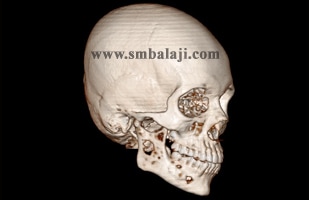
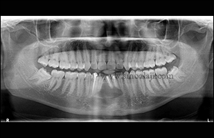
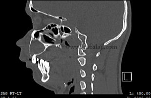
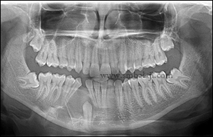
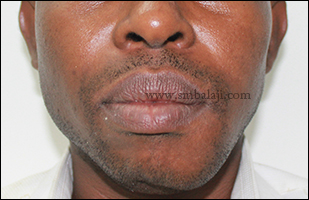
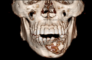
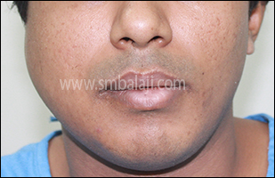
​30 year old women reported to our hospital with the complaints of swelling in right side lower half of the face. She also said that this swelling was there for a very long period with occasional numbness in the right side of the face.
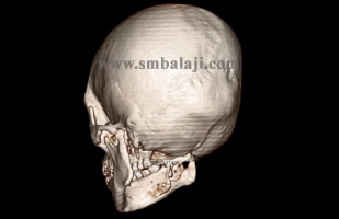
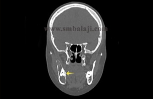
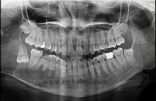
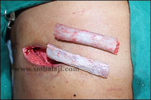
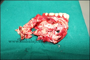
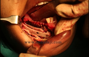
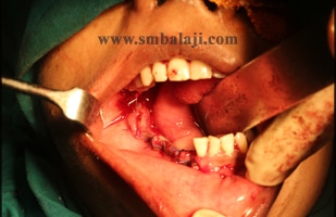
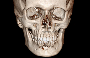
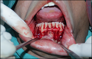
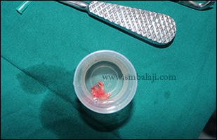
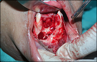
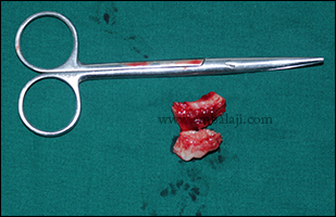
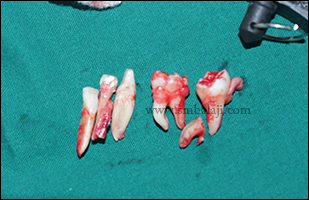
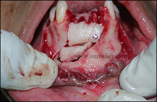
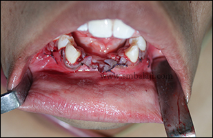
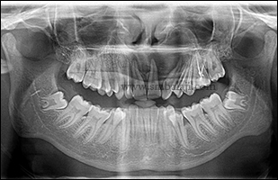
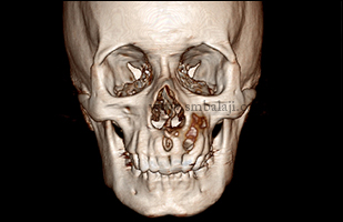
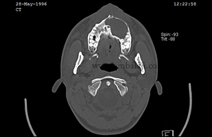
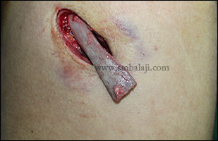
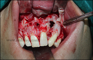
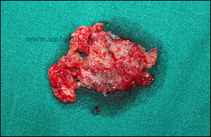
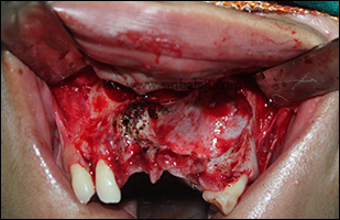
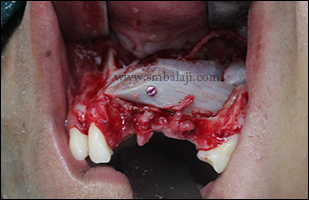
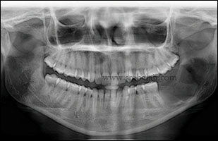
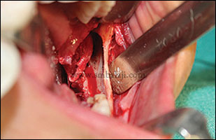
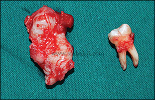
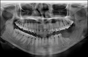
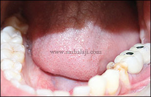
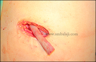
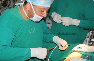
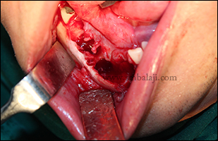
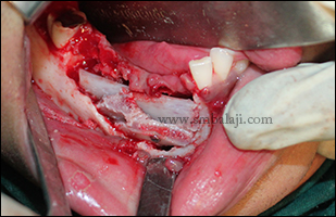
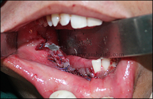
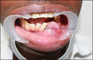
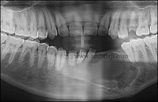
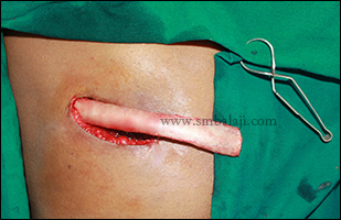
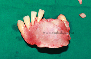
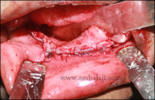
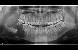
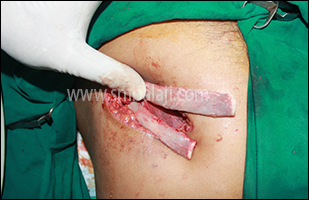
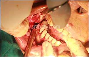
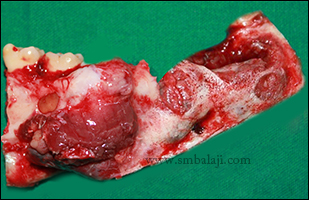
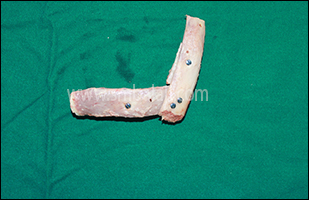
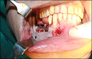
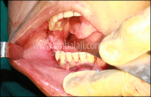
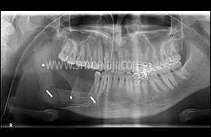
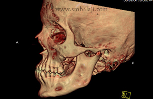
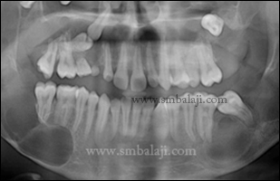
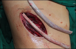
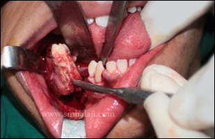
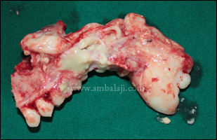
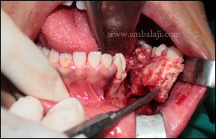
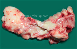
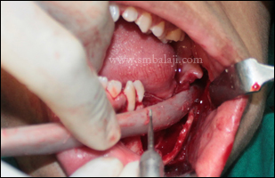
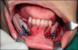
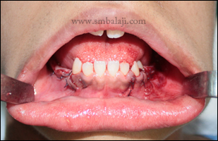
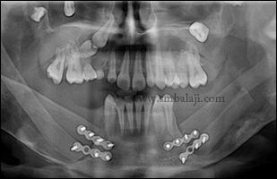
After thorough clinical, radiological and histopathological examination, Maxillofacial Surgeon Dr. S. M. Balaji diagnosed it as odontogenic keratocyst (OKC) involving right body and ramus of the mandible. He planned to remove the entire lytic lesion and reconstruction of the affected portion of the mandible in the same surgery. Costochondral graft was obtained from the ribs to reconstruct the mandible. Through intraoral approach, after raising gingivomucoperiosteal flap extending distally, the entire lesion along with the affected portion of mandible was removed in toto. Harvested rib graft was used to reconstruct the defective region of mandible. Patient is very happy to have both removal of the entire lesion and reconstruction of the affected portion of mandible in the same surgery.
Preoperative 3DCT scan view showing erosion of the cortical bone with expansion of the body and ramus of mandible
Preoperative 3DCT scan view showing erosion of the cortical bone with expansion of the body and ramus of mandible - lingual side
Preoperative 3DCT coronal slice view showing impacted wisdom tooth with huge osteolytic lesion of left ramus of mandible
Preoperative digital orthopantomogram showing large osteolytic lesion involving entire right half of the mandible
Harvested costochondral graft used to reconstruct the mandible
Affected portion of jaw showing complete osteolysis with wide spread was removed in toto
Harvested costochondral graft used to reconstruct the huge defect
Immediately after suturing
Postoperative digital orthopantomogram showing complete reconstruction of the right side of mandible
0 Comments

Successful removal and reconstruction of cystic lesion in lower jaw 

20/8/2016

0 Comments

 
​A 28 year-old man came to our hospital with a complaint of pain in his lower jaw. A digital X-ray and 3DCT scan was taken revealed a large cystic lesion extending from canine to canine of the mandible with complete erosion of the labial plate.
 Maxillofacial surgeon Dr. S.M. Balaji expertly removed the entire cystic lesion along with the lining with affected teeth in toto. Reconstruction of the bone defect was done using bone graft obtained from the retromolar region.  Following surgery the wound healed well and the patient was happy to be cured of his ailment.
Preoperative digital orthopantomogram showing osteolytic cystic lesion involving lower anterior region of mandible
3DCT view showing buccal cortex erosion in lower anterior region of mandible
Intraoperative view showing surgical exposure of cystic lesion
Cystic lesion removed along with the lining and prepared for HPE
Bone defect after removal of the cystic lesion
Bone graft obtained from the impacted teeth region
Removed of affected mandibular anterior teeth and impacted third molars
Bone graft fixed into the defective region of mandible
Immediately after suturing
0 Comments

Complete removal of huge infected cyst involving entire anterior maxilla followed by reconstruction

26/6/2016

0 Comments

 
A 26 year-old boy form West Bengal came to our hospital with a complaint of swelling with occasional pain in his upper jaw. A digital X-ray took revealed a large cystic lesion extending anterior teeth to premolar of the maxilla with complete erosion of the labial plate.
​
Histopathological examination of the specimen showed it to be a huge infected dental cyst due to continuous trauma from occlusion resulting from anterior deep bite. Maxillofacial surgeon Dr. S. M. Balaji successfully removed the entire cystic lesion along with the lining in toto. Followed by defective region of the upper jaw was reconstructed using costochondral rib graft. Following surgery the wound healed well and the patient was happy to be cured of his ailment. After complete bone formation, fixed dental replacement of the missing teeth will be done using dental implants. 
Preoperative digital orthopantomogram showing huge osteolytic lesion in anterior region of maxilla
Preoperative 3DCT view showing huge osteolytic lesion involving entire anterior and extending to the molar region of upper jaw
Axial view showing expansile osteolytic lesion involving entire left side of maxilla
Sagittal view showing buccopalatal expansion of cyst with bodily displacement of involving teeth
Costochondral graft obtained from 7th rib to reconstruct the defective portion of upper jaw
After raising gingivomucoperiosteal flap elevated and cystic lesion exposed
Removal of infected cyst in toto
After removal of cyst bone cavity debrided and diathermy applied
Costochondral graft used to augment the defective portion of upper jaw
Immediately after suturing
0 Comments

Successful removal of huge dentigerous cyst involving entire left side of lower jaw without any scars

2/6/2016

0 Comments

 
​A 25-year-old woman reported to our hospital with a complaint of swelling with pain in her left side of lower jaw. A digital X-ray took revealed a large cystic lesion extending entire ramus and angle and part of the body of the mandible involving the molar teeth.
Histopathological examination of the specimen showed it to be a chronic infected dentigerous cyst. Maxillofacial surgeon Dr. S.M. Balaji expertly removed the entire cystic lesion along with the lining with affected teeth in toto. Following surgery the wound healed well and fixed molar teeth given using dental implants in the newly formed bone with good retention, patient was happy to be cured of her ailment and to have fixed teeth.
Preoperative intraoral view
Preoperative digital orthopantomogram showing huge osteolytic lesion involving left side of mandible
Intraoperative view showing complete distruction of the bone due to cystic lesion
Removal of cyst lesion along with the involved tooth in toto
Postoperative digital orthopantomogram showing complete removal of cyst and new bone formation
Dental implants fixed in the newly formed bone with good retention
Digital orthopantomogram showing implants fixed in newly formed bone
Cosmetically well blended fixed ceramic prosthesis loaded on to the dental implants
0 Comments

Successful removal of huge dentigerous cyst in lower jaw along with the impacted canine tooth

28/4/2016

0 Comments

 
A 23 year-old boy came to our hospital with a complaint of swelling with pain in his lower jaw. A digital X-ray took revealed a large cystic lesion extending from anterior teeth to premolar of the mandible with complete erosion of the labial plate and impacted canine tooth.
 Histopathological examination of the specimen showed it to be a dentigerous cyst. Maxillofacial surgeon Dr. S.M. Balaji expertly removed the entire cystic lesion along with the lining with affected teeth in toto. Following surgery the wound healed well and the patient was happy to be cured of his ailment.
Huge osteolytic lesion involving the lower anterior region of jaw with impacted canine tooth
Costochondral rib graft obtained to correct the defective portion of the lower jaw
Dr. S. M. Balaji operating on the patient
Intraoperative view showing complete removal of cyst along with the impacted canine tooth
Defective portion of the lower jaw was reconstructed using costochondral rib graft
Immediately after suturing
0 Comments

Successful removal of huge recurrent infected follicular cyst and reconstruction of lower jaw without any scars

15/2/2016

0 Comments

 
A 28 year old man from Nigeria came to our hospital for expert treatment for his lower jaw defect. He gave the history of surgical removal of cystic lesion which was histopathologically diagnosed as follicular cyst in his lower jaw twice in his hometown and reconstructed with Bio-oss synthetic bone protein. Recurrence of the cystic lesion noted and the swelling was initially small and gradually increased to its present size for a period of 5 months. He was worried for the recurrence and wants to have permanent solution for the same.
Maxillofacial Surgeon Dr. S.M. Balaji performed the clinical and radiological investigations. OPG and 3D CT scan showed a large radiolucent lesion extending from the lower left first molar teeth till right first premolar region. Almost entire bone in this portion of the lower jaw was lost.
​
Dr. Balaji successfully removed the cyst completely along with the affected teeth and bone. Rib graft was harvested and the used to reconstruct the jaw bone defect & the surgical site was closed. After subsequent healing, implants & ceramic crowns will be placed for fixed replacement of lost teeth.
Facial view showing swelling in the lower half of the face on the left side
Preoperative intraoral view showing huge swelling with cortical expansion and dislodged teeth in the affected region of mandible
3DCT scan view showing complete erosion of the buccal cortex and adjacent teeth root resorption due to cystic lesion
Preoperative digital orthopantomogram showing huge radiolucent lesion involving the symphysis and parasymphysis region of the mandible on the left side
Costochondral rib graft harvested to reconstruct the affected portion of mandible
Intraoperative view showing removal of the cyst along with the affected bone
Cystic lesion with affected teeth and bone removed in toto
Harvested rib graft used to reconstruct the defective portion of mandible
Immediately after suturing
0 Comments

Successful jaw reconstruction for Bangladeshi man suffering from extensive jaw cyst

9/2/2016

0 Comments

 
A 30-year-old man from Bangladesh was suffering from severe pain and swelling in his right side of lower jaw. He also complained of difficulty to open his mouth wide and bite and chew foods. He was advised by his family doctor in his home town to go to Balaji Dental and Craniofacial Hospital at Chennai, India, for specialized treatment.
Maxillofacial Surgeon Dr. S.M. Balaji thoroughly evaluated his jaw defect clinically and using advanced 3D CT scan. The scan images showed that he had multiple and extensive areas of bone loss that appeared to resemble large cystic tumors involving the almost entire right side of lower jaw.
Biopsy of the lesion was done which proved it to be odontogenic keratocysts. Dr. Balaji planned a complete reconstruction of the lower jaw right side, using the man’s own rib graft. The affected portion of the lower jaw bone was surgically removed. Diseased portions of the bone were completely and thoroughly removed to ensure that there is no recurrence. A costochondral cartilage graft (rib graft) was taken and used to reconstruct the lower law. Following a few months time, rehabilitation will be completed with implants and ceramic prosthesis. The man feels very happy for the surgery outcome and the entire surgery was done intraorally avoiding external scar formation.   
0 Comments

Successful jaw reconstruction for young boy suffering from extensive jaw cyst

14/10/2015

0 Comments

 
A 14-year-old boy from Bangladesh was suffering from severe pain in his lower teeth and lower jaw. He also complained of difficulty to bite and chew foods. His parents, both doctors, were advised by their family doctor in their home town to go to Balaji Dental and Craniofacial Hospital at Chennai, India, for specialized treatment for their son.
Maxillofacial Surgeon Dr. S.M. Balaji thoroughly evaluated his jaw defect clinically and using advanced 3D CT scan. The scan images showed that he had multiple and extensive areas of bone loss that appeared to resemble large cystic tumors involving the almost entire lower jaw. Few teeth were missing and there were several supernumerary teeth embedded inside the jaw bone.
Biopsy of the lesion was done which proved it to be odontogenic keratocysts. Dr. Balaji planned a complete lower jaw reconstruction using the boy’s own rib graft. The affected portions of the lower jaw bone on both sides were surgically removed. Diseased portions of the bone were completely and thoroughly removed to ensure that there is no recurrence. A costochondral cartilage graft (rib graft) was taken and used to reconstruct the entire lower law. Following a few months time, rehabilitation will be completed with implants and ceramic prosthesis.
The boy and his parents are very happy with the surgery outcome.  The Hon’ble Former Chief Justice of Madras High Court and Judge Supreme Court of India His Excellency H L Gokhale and his wife visited the hospital and greeted the boy and his parents. Chief Justice expressed his wishes and fed the boy a piece of cake. He also commended Dr. Balaji on his remarkable expertise and applauded his excellence and outstanding achievements in successfully performing many such complex and rare reconstructive surgeries.
Preoperative 3DCT scan view showing cystic lesion involving the left ramus and angle of mandible
Cystic lesion with cortical erosion seen in the right side of the lower jaw
X-ray showing multiple cystic tumors of jaws
Graft harvested from rib
Tumor portion of right lower jaw excised
Excised portion of left lower jaw
Left lower jaw affected with cystic tumor surgically removed
Excised portion of right lower jaw
Rib graft used to reconstruct resected portion of jaw
Complete lower jaw reconstruction using the grafts
Immediately after lower jaw reconstruction surgery
Postoperative digital orthopantomogram showing complete removal and reconstructed using costochondral rib graft with good bone formation
The Hon'ble Former Chief Justice of Madras High Court and Judge Supreme Court of India His Excellency H L Gokhale wished the boy
His Excellency fed the boy a piece of cake and wished him
0 Comments

    Categories

    All
    Abbe Flap Technique
    Advanced Transport Distraction Osteogenesis
    Advancement Genioplasty
    Alveolar Cleft Repair
    Alveolar Distraction
    Ameloblastoma
    Augmentation Genioplasty
    Augmentation Rhinoplasty
    Bat Ear Correction
    Bilateral Cleft Lip Palate
    Blepharoplasty
    Bone Defect Reconstruction
    Bone Grafting
    Bone Morphogenetic Protein(BMP)
    Broad Flattened Nose Correction
    Bulky Nose Correction
    Chin Correction
    Chin Correction Surgery
    Cleft Lip & Palate Surgery
    Cleft Nose
    Cleft Palate Repair
    Cleft Rhinoplasty
    Complete Facial Rehabilitation
    Complex Facial Fractures
    Condylar Fracture Surgery
    Correction Of Hypoplastic Ramus
    Corrective Jaw Surgery
    Cosmetic Cleft Nose Correction
    Cosmetic Nose Correction
    Cosmetic Rhinoplasty
    Cosmetic Surgery
    Craniofacial Deformities
    Craniofacial Reconstruction
    Craniofacial Surgery
    Cyst Removal
    Dental Implant
    Dentigerous Cyst
    Deviated Nose Correction
    Diplopia
    Distraction Osteogenesis
    Ear Correction
    Ear Defect
    Ear Reconstructive Surgery
    Epiphora
    Extruding Infected MEDPOR
    Eyebrow Correction
    Face Enhancement
    Face Reconstruction
    Facial Asymmetry Correction
    Facial Bone Fracture
    Facial Burn Injury
    Facial Palsy Surgery
    Fibrous Dysplasia
    Flat Nose Correction
    Forehead Bone Fracture
    Forehead Deformity
    Forehead Flap
    Frontal Bossing Correction
    Gummy Smile
    Hemifacial Microsomia
    Hypertelorism Surgery
    Implant
    Incomplete Cleft Lip
    Isolated Cleft Palate Repair
    Jaw Correction Surgery
    Jaw Cyst
    Jaw Deformity
    Jaw Fracture Surgery
    Jaw Joint Ankylosis
    Jaw Reconstructive Surgery
    Jaw Surgery
    Lip Correction
    Lip Defect Correction
    Lip Revision
    Locked Jaw
    Lower Jaw Fracture
    Low Lying Upper Eyelid Correction
    Macrostomia Correction
    Mandible Correction
    Mandible Reconstruction
    Maxillofacial Surgery India
    Maxillomandibular Distraction
    Maxilofacial Surgery
    Micrognathic Mandible Correction
    Microtia
    Nasal Glial Heterotopia
    Naso-orbito-ethmoidal Fracture
    Nose And Lip Correction
    Nose Asymmetry Correction
    Nose Correction
    Nose Job
    Nose Reconstructive Surgery
    Oral Submucous Fibrosis
    Oral Tumor Removal
    Orbital Correction
    Orehead Defect
    Orthognathic Surgery
    Parrot-beak Nose
    Parry Romberg Syndrome
    Pharyngoplasty
    Plastic Surgery
    Primary Cleft Lip Repair
    Profile EnhancemeP
    Reduction Glossectomy
    Reduction Rhinoplasty
    Removal Of Capillary Hemangioma
    RhBMP 2
    RhBMP-2
    Rhinoplasty
    Road Accident Trauma
    Road Accident - Trauma
    Scar Revision
    Secondary Cleft Palate
    Sinus Lift
    Smile Makeover
    Speech Correction
    Square-face Correction
    Surgery Without Scar
    Tessier Nose Defect
    Tissue Expansion
    TMJ Ankylosis
    Tongue Correction
    Tongue Surgery
    Trauma
    Trauma Treatment
    Triangular Shaped Forehead
    Tumor Resection
    Unilateral Cleft Lip
    Unilateral Cleft Lip And Palate
    Unilateral Cleft Lip Repair
    Velopharyngeal Incompetence

    Archives

    April 2021
    March 2021
    January 2021
    December 2020
    April 2020
    March 2019
    February 2019
    January 2018
    November 2017
    October 2017
    September 2017
    June 2017
    May 2017
    April 2017
    March 2017
    January 2017
    December 2016
    October 2016
    September 2016
    August 2016
    July 2016
    June 2016
    May 2016
    April 2016
    March 2016
    February 2016
    January 2016
    December 2015
    October 2015
    September 2015
    August 2015
    July 2015
    June 2015
    May 2015
    April 2015
    March 2015
    February 2015
    January 2015
    December 2014
    November 2014
    October 2014
    September 2014
    August 2014
    July 2014
    June 2014
    May 2014
    April 2014
    March 2014
    February 2014
    January 2014
    December 2013
    November 2013

    RSS Feed

Copyright © 2020 Balaji Dental & Craniofacial Hospital | All rights reserved | Powered by BALAJI DENTAL HOSPITAL