Best Dental Clinic in Chennai : Best Dental Implant Center in India
FOLLOW US
  • HOME
  • Surgery of The week
  • Clinical Updates
  • Archives
  • Latest News
  • VIDEOS
  • CONTACT US

Microtia ear deformity correction

16/4/2020

0 Comments


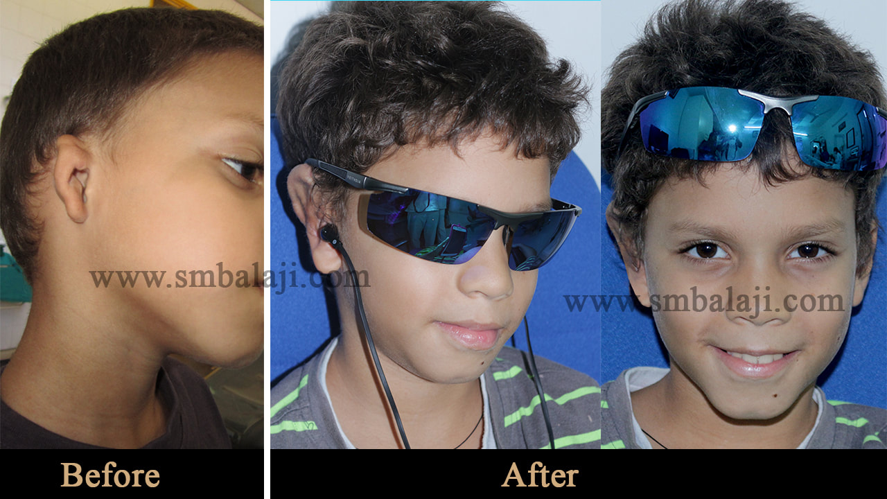
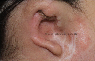
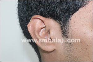
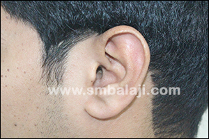
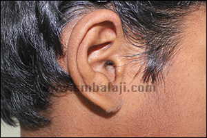
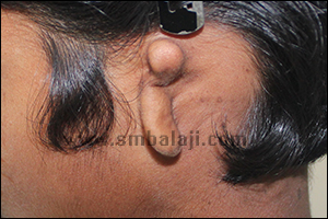
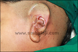
Patient born with a congenital right microtia ear deformity

​The patient is a 14-year-old boy who was born with a deformed right external ear. There was just a rudimentary structure in place. He had faced significant bullying in school. One of his greatest wishes in life was to be able to wear sunglasses like his father. His parents’ widespread enquiries for the best surgeon to address this issue led them to our hospital.

Treatment planning explained to the parents in detail

​Dr SM Balaji, microtia ear deformity surgeon, examined the patient and obtained detailed measurements of his normal left ear. He explained to the parents that the surgery needed to be performed in two stages. It was further explained that cartilaginous grafts needed to be harvested for the procedure. The parents consented to the procedure

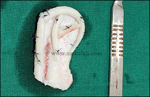
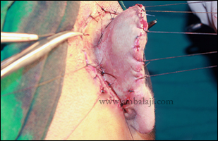
First stage of cartilaginous recreation of external ear structure

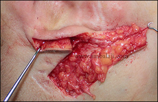
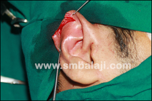
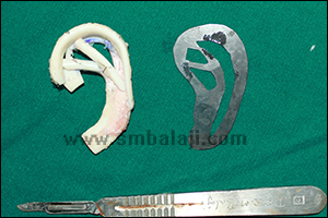
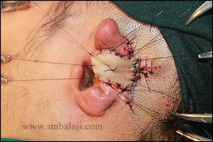
​Under general anesthesia, a costochondral graft with an intact synchondrosis was first harvested from the patient. This was in the form of an ‘E’ in order to recreate the cartilage of the external ear. Using a template created from the measurements obtained from the normal ear, the graft was carved to recreate the external ear form.
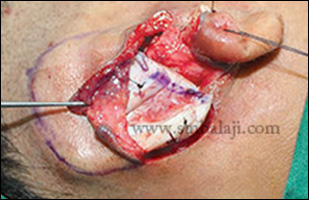
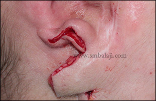
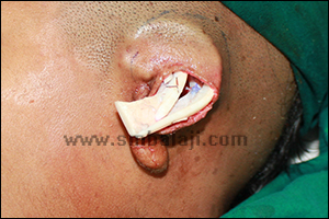
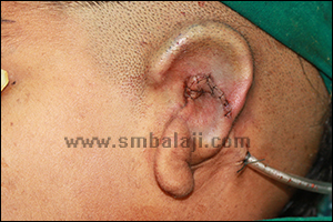
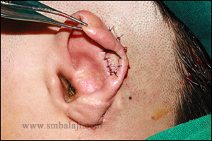
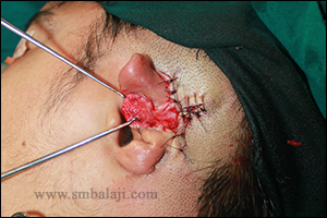
Attention was next turned to placement of the costochondral graft at the site of the deformed right ear. A subcutaneous pocket was created followed by insertion of the graft into the pocket. The incision was closed and a period of three months was allowed to elapse before the second stage.
Picture

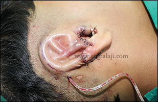
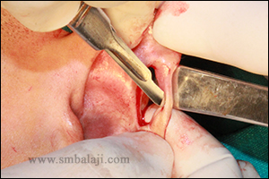
Second stage of raising the cartilaginous graft to recreate the perfect external ear

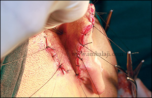
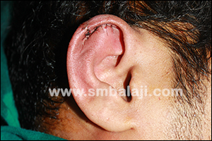
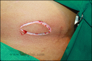
​A full thickness skin graft was harvested from the patient’s inguinal (groin) region. This was followed by incising around the cartilaginous graft and raising up the entire superstructure of the recreated right ear. The skin graft was then sutured to cover the exposed tissue behind the recreated pinna. A period of one month was allowed for full healing of the surgical site.

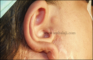
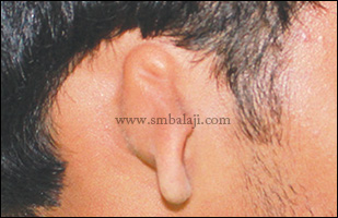
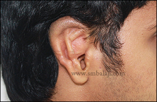
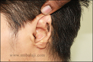
Patient expresses complete satisfaction at the results of the surgery

​The patient and his parents were very happy with the results of the surgery. He was now able to realize his long-cherished dream of wearing sunglasses like his father. His earlobe had also been perfectly recreated. They expressed their profuse gratitude to the surgical team before final discharge from the hospital.

Surgery Video

Microtia ear deformity correctionhttps://t.co/tq7u0ocdW0#Microtia #EarReconstruction #DrSMBALAJI #BalajiDental pic.twitter.com/4EYmcFANrU

— Balaji Dental and Craniofacial Hospital (@balajidental) April 16, 2020
0 Comments

Ear reconstruction for microtia 

4/6/2016

0 Comments

 
A 24-year old female from Mumbai born with a deformed right ear, sought the expertise of Maxillofacial Surgeon Dr. S.M. Balaji for reconstruction of her ear defect. Previously, in some other hospital elsewhere, multiple attempts to reconstruct the ear were done but she was not satisfied with the results. Hearing was normal.
Dr. Balaji employed the Limberg’s flap to successfully reconstruct the ear lobule. Followed which stage wise complete ear lobe reconstruction done using costochondral rib graft. Costal cartilage was harvested; external framework was carved and tunneled to reconstruct the external ear.
 After suction, the skin was seen getting adhered to the cartilage.  This was followed by the second-stage surgery to elevate the newly formed ear from the side of the head using split skin graft obtained from right thigh region. Thus, entire shape and size of the ear was reconstructed with enhanced appearance without much visible scars.
Appearance of right ear deformity
Cartilage graft harvested and carved to form ear framework
Framework placed beneath the skin to reconstruct the ear
Limberg flap raised
Earlobe reconstruction using Limberg flap
Immediately after suturing
Skin graft used to complete the elevation giving the ear a normal, raised appearance
Postoperative view following complete ear reconstruction giving enhanced appearance
0 Comments

Surgical correction of defective helix of the ear

8/2/2016

0 Comments

 
25 year old man reported to our hospital seeking for cosmetic correction of his right ear defect. He consulted many plastic surgeons for the surgical correction but was not satisfied.
On examination, the helix of the right ear was defective. Maxillofacial Surgeon Dr. S. M. Balaji successfully corrected his ear defect using wedge closure technique without much visible scars. Results are immediate. He is very happy to have single stage ear defect correction. 
0 Comments

Microtia - Ear Creation/Reconstruction Surgery - Dr. S.M Balaji

31/1/2016

0 Comments

 
Microtia is a congenital malformation of variable severity of the external and middle ear. This condition is corrected by staged ear reconstruction using autogenous costal cartilage.
Shown here is a case of pediatric external ear reconstruction. The girl’s left microtic auricle had remnants of disorganized cartilage with an earlobe. This surgery is normally performed at about 14-15 years of age but it was done at a younger age in this 10-year-old because she was facing psychological problems as a result of being teased a lot for her ear defect by other children at school.
A template of the normal side was designed to recreate the key elements of the ear shape on the affected side. Costal cartilage was harvested; external framework was carved and tunneled to reconstruct the external ear. After suction, the skin is seen getting adhered to the cartilage.  This will be followed by the second-stage surgery to elevate the newly formed ear from the side of the head.
Ear reconstruction surgeries are a demanding undertaking and must be performed with skill, precision & artistry.
Normal right ear
Microtia left ear
Intraoperative view - using template rib cartilage harvested
Harvested cartilage shaped in the form of ear
Banking of the cartilage beneath the skin
Cartilage in position
Dr S M Balaji performing the ear reconstruction surgery
Immediately after suturing showing reconstructed ear lobe with suction drain in position
0 Comments

Ear reconstruction for Microtia (ear deformity)

18/6/2015

0 Comments

 
Microtia (ear deformity) of right ear
Cartilage graft harvested and carved to form ear framework
Suction drain placed for skin to adhere to cartilage framework
Skin graft used to complete the elevation giving the ear a normal, raised appearance
More normal appearance and improved aesthetics following ear reconstruction
A young boy was born with a deformity of his right ear. The external portion of his right ear was deformed due to which he suffered ridicule and unwelcome stares. He was very depressed about his appearance. His parents were very worried about their son’s psychological well-being. Seeking the best treatment they brought him to our hospital. Maxillofacial Surgeon Dr. S.M. Balaji successfully performed a staged ear reconstruction surgery to correct the microtia defect.  On examination it was seen that the right ear was smaller in size than the left ear with absence of helix, antihelix, scapha. The external opening to the ear canal was decreased in size and there was no associated deafness. The lobule was present and inferiorly placed. For the reconstruction, a costochondral graft (rib graft) was harvested. Based on the measurements of the normal left ear, the graft was carved and chiseled to form a framework.  The skin of the defective ear was elevated and the cartilage framework was placed in the pocket. A suction drain was placed for the skin to adhere to the cartilage. In the second stage surgery, the newly formed ear was elevated from the side of the head using a cartilage and skin graft. Following reconstruction the ear was given a more normal, raised appearance. The boy is happy that he won’t be teased any longer and he and his parents were very happy with the surgery outcome.

Ear reconstruction surgeries are a demanding undertaking and must be performed with skill, precision & artistry.

0 Comments

Successful reconstruction surgery for ear defect

2/6/2014

0 Comments

 
An 18 year old boy was brought to our hospital by his parents for complete reconstruction of his ear defect. He had a large button-holing defect in the helix of his left ear due to which he was teased a lot and it affected his self-confidence.

Maxillofacial Surgeon Dr. S.M. Balaji skillfully performed the surgical ear reconstruction in 2 stages. In the first stage the specialized island flap was raised just behind the ear and the edges of the button holing defect in the pinna of the ear was cut. The defective pinna of the ear was sutured with the island flap behind the ear. In the second stage after 6 months, the pinna of the ear was separated from the flap behind the ear and the detached region was sutured. The size of the defect was measured using lint and the required size of skin graft was harvested to close the raw plane of the ventral surface of the ear and it was sutured by means of quilting technique to avoid spacing between the skin graft and recipient site and seroma formation. Thus total reconstruction was done without much surgical scars. Results are immediate and we will update this case by post operative photos with complete healing later. 
Pre operative view showing button hole defect in pinna of the left ear
The defective region was surgically operated by placing a cut in the margins
Region just behind the ear was marked and flap raised and the pinna of the ear was attached and sutured
In the second stage the pinna of the ear was surgically lifted and the excised region was sutured
Skin graft of exactly same size of the defect was harvested from the hip region
Skin graft was sutured to the pinna of the ear by quilting technique
0 Comments

    Categories

    All
    Abbe Flap Technique
    Advanced Transport Distraction Osteogenesis
    Advancement Genioplasty
    Alveolar Cleft Repair
    Alveolar Distraction
    Ameloblastoma
    Augmentation Genioplasty
    Augmentation Rhinoplasty
    Bat Ear Correction
    Bilateral Cleft Lip Palate
    Blepharoplasty
    Bone Defect Reconstruction
    Bone Grafting
    Bone Morphogenetic Protein(BMP)
    Broad Flattened Nose Correction
    Bulky Nose Correction
    Chin Correction
    Chin Correction Surgery
    Cleft Lip & Palate Surgery
    Cleft Nose
    Cleft Palate Repair
    Cleft Rhinoplasty
    Complete Facial Rehabilitation
    Complex Facial Fractures
    Condylar Fracture Surgery
    Correction Of Hypoplastic Ramus
    Corrective Jaw Surgery
    Cosmetic Cleft Nose Correction
    Cosmetic Nose Correction
    Cosmetic Rhinoplasty
    Cosmetic Surgery
    Craniofacial Deformities
    Craniofacial Reconstruction
    Craniofacial Surgery
    Cyst Removal
    Dental Implant
    Dentigerous Cyst
    Deviated Nose Correction
    Diplopia
    Distraction Osteogenesis
    Ear Correction
    Ear Defect
    Ear Reconstructive Surgery
    Epiphora
    Extruding Infected MEDPOR
    Eyebrow Correction
    Face Enhancement
    Face Reconstruction
    Facial Asymmetry Correction
    Facial Bone Fracture
    Facial Burn Injury
    Facial Palsy Surgery
    Fibrous Dysplasia
    Flat Nose Correction
    Forehead Bone Fracture
    Forehead Deformity
    Forehead Flap
    Frontal Bossing Correction
    Gummy Smile
    Hemifacial Microsomia
    Hypertelorism Surgery
    Implant
    Incomplete Cleft Lip
    Isolated Cleft Palate Repair
    Jaw Correction Surgery
    Jaw Cyst
    Jaw Deformity
    Jaw Fracture Surgery
    Jaw Joint Ankylosis
    Jaw Reconstructive Surgery
    Jaw Surgery
    Lip Correction
    Lip Defect Correction
    Lip Revision
    Locked Jaw
    Lower Jaw Fracture
    Low Lying Upper Eyelid Correction
    Macrostomia Correction
    Mandible Correction
    Mandible Reconstruction
    Maxillofacial Surgery India
    Maxillomandibular Distraction
    Maxilofacial Surgery
    Micrognathic Mandible Correction
    Microtia
    Nasal Glial Heterotopia
    Naso-orbito-ethmoidal Fracture
    Nose And Lip Correction
    Nose Asymmetry Correction
    Nose Correction
    Nose Job
    Nose Reconstructive Surgery
    Oral Submucous Fibrosis
    Oral Tumor Removal
    Orbital Correction
    Orehead Defect
    Orthognathic Surgery
    Parrot-beak Nose
    Parry Romberg Syndrome
    Pharyngoplasty
    Plastic Surgery
    Primary Cleft Lip Repair
    Profile EnhancemeP
    Reduction Glossectomy
    Reduction Rhinoplasty
    Removal Of Capillary Hemangioma
    RhBMP 2
    RhBMP-2
    Rhinoplasty
    Road Accident Trauma
    Road Accident - Trauma
    Scar Revision
    Secondary Cleft Palate
    Sinus Lift
    Smile Makeover
    Speech Correction
    Square-face Correction
    Surgery Without Scar
    Tessier Nose Defect
    Tissue Expansion
    TMJ Ankylosis
    Tongue Correction
    Tongue Surgery
    Trauma
    Trauma Treatment
    Triangular Shaped Forehead
    Tumor Resection
    Unilateral Cleft Lip
    Unilateral Cleft Lip And Palate
    Unilateral Cleft Lip Repair
    Velopharyngeal Incompetence

    Archives

    April 2021
    March 2021
    January 2021
    December 2020
    April 2020
    March 2019
    February 2019
    January 2018
    November 2017
    October 2017
    September 2017
    June 2017
    May 2017
    April 2017
    March 2017
    January 2017
    December 2016
    October 2016
    September 2016
    August 2016
    July 2016
    June 2016
    May 2016
    April 2016
    March 2016
    February 2016
    January 2016
    December 2015
    October 2015
    September 2015
    August 2015
    July 2015
    June 2015
    May 2015
    April 2015
    March 2015
    February 2015
    January 2015
    December 2014
    November 2014
    October 2014
    September 2014
    August 2014
    July 2014
    June 2014
    May 2014
    April 2014
    March 2014
    February 2014
    January 2014
    December 2013
    November 2013

    RSS Feed

Copyright © 2020 Balaji Dental & Craniofacial Hospital | All rights reserved | Powered by BALAJI DENTAL HOSPITAL