Best Dental Clinic in Chennai : Best Dental Implant Center in India
FOLLOW US
  • HOME
  • Surgery of The week
  • Clinical Updates
  • Archives
  • Latest News
  • VIDEOS
  • CONTACT US

Cleft rhinoplasty without scars - BALAJI DENTAL

27/6/2015

Comments

 
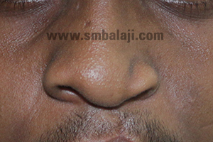
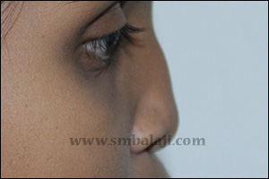
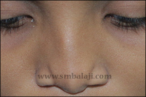
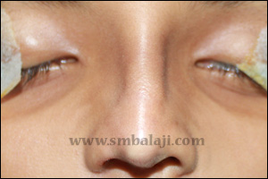
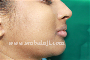
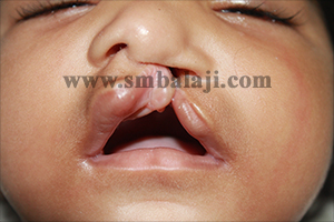
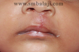
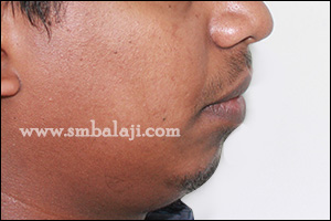
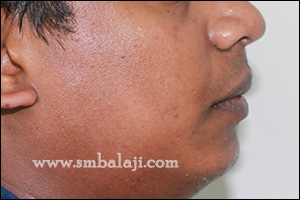
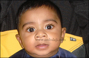
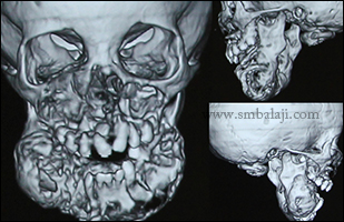
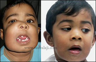
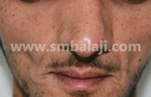
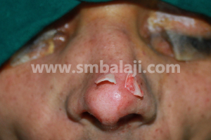
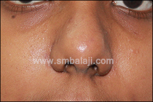
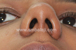
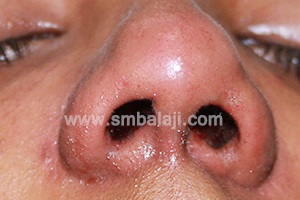
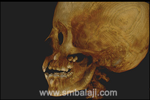
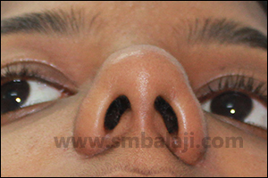
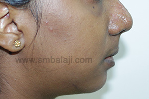
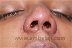
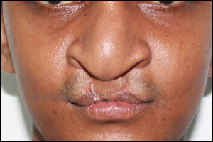
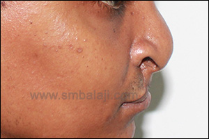
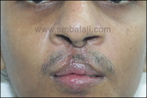
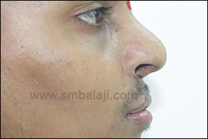
A 22-year-old boy reported to our hospital seeking specialized treatment to correct his cleft nose defect. He was previously operated for cleft lip and palate elsewhere in his childhood. Due to the cleft defect his nose was collapsed on the left side and it affected his facial appearance.
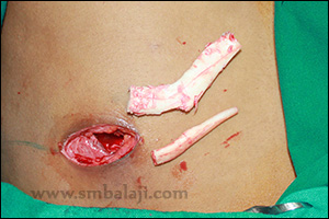
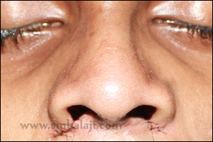
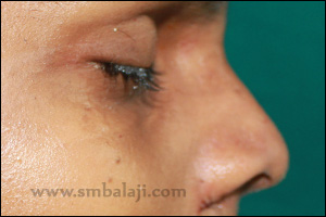
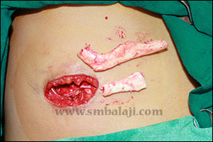
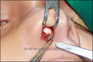
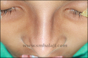
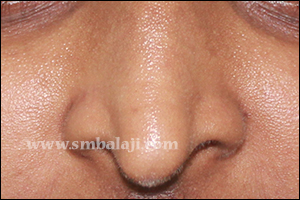
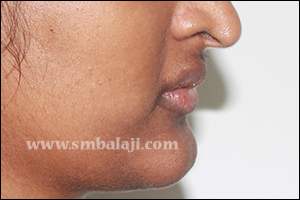
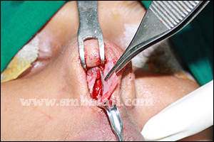
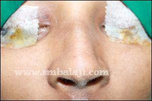
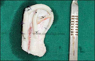
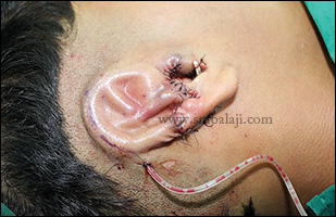
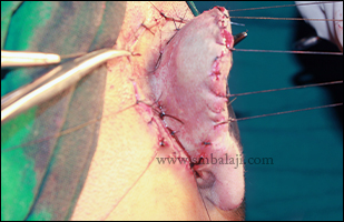
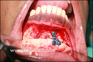
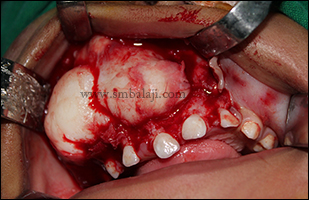
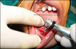
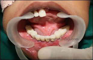
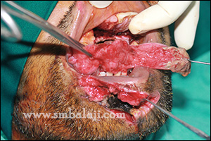
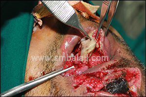
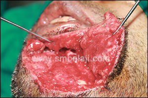
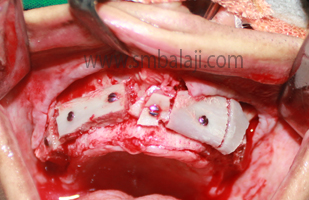
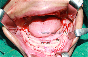
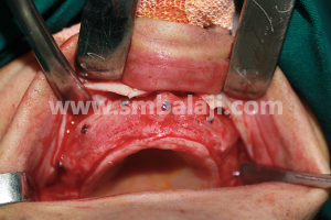
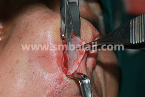
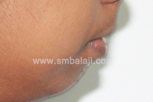
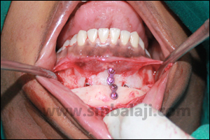
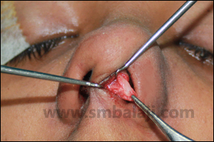
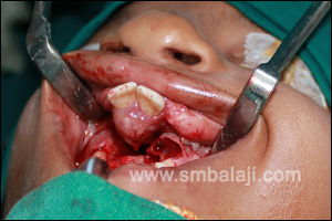
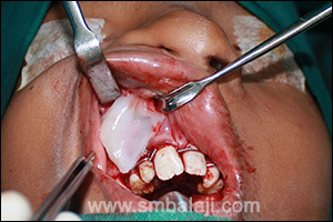
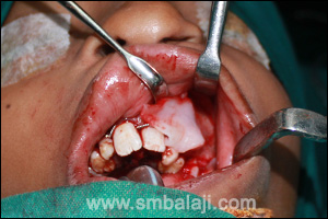
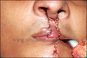
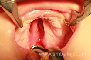
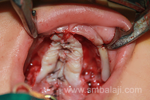
Maxillofacial Surgeon Dr. S.M. Balaji expertly performed the rhinoplasty or nose correction. A costochondral graft (rib graft) was harvested and used to reconstruct the collapsed nasal framework. The surgery was approached from inside the nose so there was no scar. Immediately after surgery, the nose appeared sharp and definite giving a more pleasing appearance. The patient was very happy to have his nose defect corrected without any scars.  
Facial view showing flat ala on the left side due to cleft defect
Flat dorsum with depressed columella due to cleft defect
Depressed columella with alar defect due to cleft deformity
Harvested costochondral rib graft used to augment the dorsum of the nose
Immediate postoperative view showing corrected alar defect
Corrected profile view showing augmented dorsum of the nose
Corrected alar shape with nostrils and columellar length raised without any visible scars on the face
Comments

Augmentation rhinoplasty for improved appearance

24/6/2015

Comments

 
An 18-year-old girl was brought to our hospital by her parents to correct the shape and appearance of her nose. Her nose was flattened and ill-defined that gave her an unattractive appearance. She was very depressed about her looks.
Maxillofacial Surgeon Dr. S.M. Balaji successfully performed the augmentation rhinoplasty. A costochondral graft was harvested from the girl’s rib and used to reconstruct the nasal dorsum. The procedure was done from inside the nose so there were no scars. Immediately following surgery the nose appeared sharp, defined and pristine that made her look more attractive. The girl and her parents were very happy with the outcome of the surgery.
Preoperative facial view showing flat dorsum of the nose
Profile view showing flat dorsum of nose with illdefined bridge of the nose
Illdefined bridge of the nose
Costochondral rib graft harvested to reconstruct the dorsum of nose
Intranasally the harvested rib graft inserted into the dorsum of the nose
Immediate postoperative view showing corrected nose profile without any visible surgical marks on the face
Augmented dorsum of the nose giving pristine appearance
Raised bridge of the nose
Comments

Combined nose and chin reduction surgery for enhanced appearance

22/6/2015

Comments

 
​A 23-year-old girl came to our hospital seeking to correct the shape of her nose and chin to improve her appearance. Her nose appeared very long & the nose tip was very drooping and her chin was disproportionate & protruding. This gave her an unattractive appearance and was making her very self-conscious.
Maxillofacial Surgeon Dr. S.M. Balaji successfully performed a combined reduction rhinoplasty and reduction genioplasty. The caudal portion of the lower lateral nasal septal cartilages were trimmed. Nose was reshaped making it sharp and pristine. Excess chin bone was also trimmed. Immediately following surgery the girl’s facial profile was enhanced and she was happy to have more attractive facial features without any scar on the face.
Preoperative facial view showing long nose with elongated tip of the nose
Preoperative profile view showing long dorsum of nose with drooping tip and prominent chin
Intraoperative view showing removal of caudal portion of nasal septal cartilage to reduce the length of the nose
Immediate postoperative view showing corrected length of the nose without any visible surgical marks on the face
Postoperative profile view showing corrected nose and chin profile giving pristine appearance
Comments

Primary lip repair for baby with unilateral cleft lip and palate

20/6/2015

Comments

 
A 3-month-old baby girl born with unilateral cleft lip and palate was brought to our hospital by her parents for specialized treatment of the defect.
Maxillofacial Surgeon Dr. S.M. Balaji successfully performed the primary cleft lip repair using modified Millard’s technique.  Right away after surgery the baby’s appearance was remarkable improved and she was able to feed well. Her parents were very happy to have the cleft lip surgically treated with negligible scar. After few months cleft palate repair will be done.
Preoperative view showing unilateral cleft lip and palate defect in 3 months old baby girl
Immediate postoperative view showing complete closure of the cleft lip defect
Immediately after suture removal following seven days postoperative view showing enhanced appearance
Comments

Ear reconstruction for Microtia (ear deformity)

18/6/2015

Comments

 
Microtia (ear deformity) of right ear
Cartilage graft harvested and carved to form ear framework
Suction drain placed for skin to adhere to cartilage framework
Skin graft used to complete the elevation giving the ear a normal, raised appearance
More normal appearance and improved aesthetics following ear reconstruction
A young boy was born with a deformity of his right ear. The external portion of his right ear was deformed due to which he suffered ridicule and unwelcome stares. He was very depressed about his appearance. His parents were very worried about their son’s psychological well-being. Seeking the best treatment they brought him to our hospital. Maxillofacial Surgeon Dr. S.M. Balaji successfully performed a staged ear reconstruction surgery to correct the microtia defect.  On examination it was seen that the right ear was smaller in size than the left ear with absence of helix, antihelix, scapha. The external opening to the ear canal was decreased in size and there was no associated deafness. The lobule was present and inferiorly placed. For the reconstruction, a costochondral graft (rib graft) was harvested. Based on the measurements of the normal left ear, the graft was carved and chiseled to form a framework.  The skin of the defective ear was elevated and the cartilage framework was placed in the pocket. A suction drain was placed for the skin to adhere to the cartilage. In the second stage surgery, the newly formed ear was elevated from the side of the head using a cartilage and skin graft. Following reconstruction the ear was given a more normal, raised appearance. The boy is happy that he won’t be teased any longer and he and his parents were very happy with the surgery outcome.

Ear reconstruction surgeries are a demanding undertaking and must be performed with skill, precision & artistry.

Comments

Chin advancement surgery (advancement genioplasty) for enhanced appearance

18/6/2015

Comments

 
A 23-year-old man came to our hospital seeking to advance his chin to improve his appearance. His chin was too retruded (set back) that gave him an unattractive facial profile making him depressed and conscious about his looks.
Maxillofacial Surgeon Dr. S.M. Balaji successfully performed the advancement genioplasty. The chin bone was sectioned, brought forward and stabilized in a more balanced position. This was done from inside the mouth itself so there were no scars. Immediately following surgery, the man’s facial profile improved considerably that boosted his self-confidence. He is very happy with the surgery results. 
Preoperative profile view showing retruded chin
Intraoperative view showing advancement genioplasty
Postoperative profile view showing enhanced chin profile giving perfect facial harmony without any scars on the face
Comments

 Single stage Removal & Reconstruction of jaw tumor in a 1 year old boy for the first time and rehabilitation with dental implants

17/6/2015

Comments

 
For the 1st time a rare type of aggressive facial and jaw bone tumor for 1 year old boy was removed through the mouth and the jaws reconstructed using rib graft, titanium mesh and bone morphogenetic protein. Rehabilitation was completed using dental implants and ceramic prosthesis to replace the missing teeth.  

A path-breaking surgery has helped an infant to recover from a facial disfigurement and life threatening ailment owing to a tumor in the jaw bone. The boy’s parents were very worried and they had approached many centers across the globe but the doctors there were hesitant to treat the one-year-old Maldivian boy due to the complexity of his condition. Dr. S.M. Balaji renowned for his expertise and dedication in Maxillofacial surgery successfully accomplished this complex task.

The Maldivian boy Saalik was 1 year old when he was referred to the hospital. He was suffering from a rare form of jaw bone disease called Osteodystrophy. It is a kind of tumor wherein the jaw bone dissolves gradually and gets replaced by soft tissues. Owing to this, his face was disfigured and his ability to eat and speak was becoming increasingly difficult. The disease severity was rapidly increasing from the time he was just 8 months of age.

Conventionally, for such cases, the only treatment is surgery which should be done when most of the growth ceases, i.e. at about 15 years of age. But in this instance, the extent of the tumor was vast involving the entire upper jaw and almost whole of lower jaw except the jaw joints. Distorted jaws will impede the growth of other facial bones also. Hence the only option was to intervene surgically immediately.

In a six-hour surgery, the diseased portions of upper and lower jaws were removed. A bone graft was taken from the boy’s rib and along with a customized imported Titanium plate was used to reconstruct the jaws. To enhance bone healing rhBMP-2 was also placed.

Recombinant human Bone Morphogenetic Protein is a scientifically prepared version of a protein that is already present in the body that promotes new bone growth. It stimulates the patients’ own cells to rapidly form new bone and quickly heals bony defects. This miracle protein also finds use in treatment of other maxillofacial defects like cysts and fractures. Other than the US, this centre is the only facility in this part of the world to adopt this technology with great success.

The widespread extent and severity of the disease and that too at such a young age complicated the problem. Microvascular surgery is tough due to the very fine blood vessel structures. Surgical strategies were planned meticulously using advanced CT scans. The innovative 3-in-1 surgery avoided the need for further surgeries. For the first time such an attempt to completely rehabilitate the tumor in a single stage was done.

The highlight of the surgery was that the entire procedure was done from inside his mouth so there will not be any visible scars on the child’s face. Following good new bone formation, his rehabilitation was completed with dental implants and fixed ceramic crown prosthesis to replace his missing teeth. He is able to eat and chew well. The boy’s parents are very happy that their child’s ailment has been successfully treated and that he is able to breathe, eat and speak well while at the same time his facial appearance is also normal again.

Saalik’s positive response and normal health have showed us how this surgery and stem cells treatment can be potentially used for reconstruction. 

Normal appearance of the baby at 6 months of age before tumor growth
Huge disfiguring facial swelling affecting the appearance and inability to close the mouth and speak properly
3DCT scan view showing complete destruction of the upper and lower jaw bone
Huge expansile bone tumor involving the entire maxilla was surgically exposed
Huge tumor was removed intraorally
Maxillary tumor mass in toto
Completely excised mandible along with the tumor mass
New bone formation following reconstruction of the jaw bone using costochondral rib graft and rhBMP2 bone protein
3DCT scan view showing good bone formation
Before and after tumor resection without any visible scars on the face
Dr. S. M. Balaji with the boy following successful jaw reconstruction
Implant fixation in the newly formed alveolar jaw bone
Dental implants fixed into the jawbone with good stability and retention
Cosmetically well blended implant prosthesis given
The boy and his parents are happy to have the tumor treated well without scar and for new teeth with implants
Comments

Necrosed split portion of tongue and chin wound sustained in road traffic accident successfully reconstructed

17/6/2015

Comments

 
A 39-year-old man sustained severe facial injuries in a road traffic accident. As a result of the trauma, a large part of the left side of his tongue was split. Also he sustained a through and through wound on his chin that led to a communication between the chin to the floor of the inner part of the lower lip (labial vestibule). This caused saliva to drool from the mouth onto the surface of the chin through the wound. He was brought to our hospital for immediate treatment.
Maxillofacial Surgeon Dr. S.M. Balaji successfully performed the reconstructive surgery. The necrosed split portion of tongue was removed and rest approximated and the musculature sutured in layers restoring the form and function of the tongue. Layer-wise closure of the chin wound was done to close the communication between the mouth and chin ensuring very negligible scar.  The patient is happy with the surgery outcome and for the surgical repair of his injuries.
Split tongue injury with severe cut wound in chin region due to an accident
Necrosed skin in the chin region and necrosed tongue portion identified and removed
Intraoperative view showing huge chin defect with removal of necrosed skin
Immediately after suturing the tongue defect and extraoral cut wounds
Following 5 days of surgery showing enhanced appearance with good approximation of tongue maintaining the function
Comments

Simultaneous upper jaw reconstruction with bone graft, implant fixation and reduction rhinoplasty

16/6/2015

Comments

 
A young man from the Middle East was referred to our hospital for fixed replacement of his multiple missing teeth (anodontia). Along with placing implants, the patient also wanted to correct his nose shape to improve his appearance. A 3D CT scan revealed extreme deficiency of bone in his upper and lower jaw insufficient for placement of implants. Maxillofacial Surgeon & Implantologist Dr. S.M. Balaji reconstructed the maxilla and mandible using bone graft taken from the rib region.

Dr. Balaji successfully placed implants with good retention and stability in the reconstructed jaws. His nose was very long, broad, crooked and misshapen giving him an unattractive appearance. Simultaneously reduction rhinoplasty was performed to correct the nose shape. Lateral osteotomy was done to correct the deviated septum. Lower lateral nasal cartilages were trimmed to improve the appearance of the nose giving it a more balanced form enhancing the appearance. The patient was very happy with the surgery outcome without any scar on the face.
3DCT scan view showing complete anodontia of both maxilla and mandible with thin alveolar process
Intraoral view showing completely edentulous maxilla and mandible
Preoperative digital orthopantamogram showing completely edentulous jaws with iildefined alveolar process in maxilla
Harvested rib graft used to augment the maxillary alveolar ridge
Dental implants fixed in the lower jaw and stabilized with rib graft for good retention
3DCT scan showing good uptake of bone graft and osseointegration of lower implants
Facial view showing long deviated nose
Good bone formation following bone grafting
Implants fixed with good stability and retention in the newly formed bone
Immediately after suturing the gingivomucoperiosteal flap
Removal of caudal portion of lateral nasal septal cartilage to corret the bulkiness in the tip of the nose
Septal deviation was corrected intranasally
Immediate postoperative view showing pristine nose without any visible surgical marks on the face
Pre and post operative nose views
Postoperative OPG showing dental implants fixed with good retention in reconstructed maxilla and mandible
Comments

Combined nose and chin correction for improved appearance

15/6/2015

Comments

 
​A 23-year-old girl came to our hospital seeking to correct her nose and chin defect. Her chin was very retruded and the tip of her nose was very bulky. She was very self-conscious about her appearance and wanted to improve her facial features.
Maxillofacial Surgeon Dr. S.M. Balaji successfully performed a combined nose and chin correction. Advancement genioplasty was done to correct the retruded chin. The chin bone was sectioned, brought forward and fixed in a more balanced position. The bulbous nose tip was corrected with reduction rhinoplasty.  Foot plate of the medial crus and lower lateral nasal cartilages were removed giving the nose a more slender and sharp. With these combined procedure the patient was very happy to have a more enhanced facial profile without any surgical scars. 
Bulky tip of the nose giving unpleasant appearance
Misshapen nose affecting the facial appearance
Profile view showing severely retruded & improperly aligned lower jaw
Advancement genioplasty - chin bone sectioned, brought forward and fixed in more balanced position
Footplate of the medial crus removed
Postoperative profile view showing augmented chin profile without any visible surgical marks on the face
Improved appearance of the nose enhancing appearance without scar
Comments

Successful union of cleft bone into a single jaw bone using miracle protein rhBMP-2

12/6/2015

Comments

 
​An 8 year old boy with bilateral cleft lip and palate defect from Bihar was brought to our hospital with his parents for closure of the cleft defect in the teeth bearing region of upper jaw bone and further rehabilitation of his cleft deformity. The primary cleft lip and palate repair was done elsewhere.
The premaxilla was separated from rest of the alveolus which can be clearly appreciated in 3D CBCT scan. Thus upper alveolus was separated into three different parts which will severely affect the development of teeth later.
Maxillofacial Surgeon Dr. S. M. Balaji performed the premaxillary setback procedure with rhBMP-2 which helps in uniting the three parts into a single jaw bone with new bone formation. This surgery helped for proper growth and development as well as alignment of his teeth.
Intraoperative view showing separate premaxilla due to bilateral cleft defect
3D CBCT scan showing detached and forwardly placed premaxilla from rest of the alveolus
Miracle bone protein rhBMP2 placed in the cleft region to induce new bone formation
Premaxillary segment aligned with the rest of the alveolus and stabilized
Immediately after suturing the gingivomucoperiosteal flap
Comments

Combined nose and chin correction for improved appearance

8/6/2015

Comments

 
A 23-year-old girl came to our hospital seeking to correct her nose and chin defect. Her chin was very retruded and the tip of her nose was very bulky. She was very self-conscious about her appearance and wanted to improve her facial features.

Maxillofacial Surgeon Dr. S.M. Balaji successfully performed a combined nose and chin correction. Advancement genioplasty was done to correct the retruded chin. The chin bone was sectioned, brought forward and fixed in a more balanced position. The bulbous nose tip was corrected with reduction rhinoplasty.  Foot plate of the medial crus and lower lateral nasal cartilages were removed giving the nose a more slender and sharp. With these combined procedure the patient was very happy to have a more enhanced facial profile without any surgical scars. 
Bulky tip of the nose giving unpleasant appearance
Misshapen nose affecting the facial appearance
Profile view showing severely retruded & improperly aligned lower jaw
Advancement genioplasty - chin bone sectioned, brought forward and fixed in more balanced position
Postoperative profile view showing augmented chin profile without any visible surgical marks on the face
Improved appearance of the nose enhancing appearance without scar
Comments

Nose correction & Abbe flap procedure for lip correction in cleft patient

8/6/2015

Comments

 
A 25-year-old man came to the hospital wanting to correct his lip and nose defect to improve his appearance. He was born with bilateral cleft lip and palate which was previously operated elsewhere. His upper lip was very thin and his nose appeared deformed making him very self-conscious about his appearance.

Maxillofacial Surgeon Dr. S. M. Balaji successfully performed an abbe flap procedure for lip correction and nose correction. A flap of tissue was taken from the lower lip, rotated across the mouth and used to reconstruct the upper lip with the base of the flap still attached to the lower lip to maintain blood supply. The flap was taken with hair follicles to ensure moustache growth. Once blood supply was established, the flap was divided giving the upper lip a more natural, improved form. The broad nose was reshaped to make it more proportionate and balanced. Following surgery the man was very happy to have an improved facial profile.
Preoperative facial view showing defective cleft nose and lip
Preoperative profile view showing depressed nasal tip due to missing columella and missing upper lip prominence
Immediately after suturing with columella enhancement and upper lip correction
Immediately after suture removal following Abbe division showing enhanced appearance of nose and upper lip without much surgical marks
Profile view showing augmented nose profile and columella and proportionate lips were obtained giving enhanced appearance
Comments

Successful isolated cleft palate repair surgery

4/6/2015

Comments

 
A 5 months old baby girl with isolated cleft palate was brought to our hospital for the surgical closure of the gap (cleft) in the roof of the mouth (palate). The girl’s cleft palate defect was classified as Millard’s 10 and 11.

Maxillofacial Surgeon Dr. S. M. Balaji surgically corrected the cleft palate using palatal pushback technique. Incision was made in the mid- vomerine region. The palatal flap was raised on both right and left side. The Levator palatine muscle was detached from their abnormal positions and reattached to its normal position like a hammock. A two layer closure was done. The nasal floor was closed with the vomerine flap making a reverse knot. Oral layer was sutured by vertical mattress sutures. Results are immediate and the suction test was positive which ensures improved speech.
Isolated cleft palate in a 5 months old baby girl
Palatal cleft closure using palatal pushback technique
Comments

Combined Hemangioma excision and chin correction surgery

3/6/2015

Comments

 
A 22-year-old girl came to our hospital seeking to treat the defect on her chin. She complained of a growth of tissue protruding on her chin which was initially small and gradually grew to the present size and the overlying skin was fiery red & blistered. This was adversely affecting her appearance. She gave a history of a similar such growth in the past in her chin which was removed elsewhere but subsequently re-occurred. She wanted a permanent treatment.

Maxillofacial Surgeon Dr. S.M. Balaji successfully performed the surgical treatment. An angiogram revealed the lesion to be a hemangioma. A 3D CT scan showed bony excess of the chin bone that gave a more protruding appearance to the chin. Dr. Balaji performed a combined genioplasty procedure removing the excess bone along with the hemagioma lesion through the scar of the previous surgery itself thereby avoiding any new scar on the face. The patient recovered well and is happy to have an improved appearance and the defect cured. 
Preoperative 3DCT scan facial view
Facial view showing hemangioma in the symphyseal region
Profile view showing prominent chin due to hemangiomatous growth along with bone protrusion
Intraoperative view showing complete removal of hemangiomatous lesion along with bone removal
Reduction genioplasty with excess bone removed in toto
After removal of excess bone adjacent segments stabilized with titanium bone plates
Immediately after suturing following excess skin removal with drain in place
Comments

Successful repair of ear tear and lower jaw fractures

1/6/2015

Comments

 
A 38-year-old man had met with a motor vehicular accident in which he had sustained severe injuries to his right ear and lower jaw. His right ear was severely torn causing him great pain and he had much difficulty opening and closing his mouth.

A 3D CT scan showed right and left body of mandible fracture. Maxillofacial Surgeon Dr. S.M. Balaji successfully performed the reconstructive surgery. The mandibular fractures were fixed with bone plates and screws. The ear tear extended from behind the ear at the side of the neck, completely splitting the pinna till the skin of the preauricular region. The scar tissues were excised; the cut pinna was closely approximated and sutured restoring normal form and function. Following suture removal the right ear had a normal appearance and the man was very happy to have his injuries treated successfully. 

Severe cut wound in right ear due to trauma
Immediately after suturing
Immediately after suture removal following seven days of surgery showing complete correction of ear
Comments

    Categories

    All
    Abbe Flap Technique
    Advanced Transport Distraction Osteogenesis
    Advancement Genioplasty
    Alveolar Cleft Repair
    Alveolar Distraction
    Ameloblastoma
    Augmentation Genioplasty
    Augmentation Rhinoplasty
    Bat Ear Correction
    Bilateral Cleft Lip Palate
    Blepharoplasty
    Bone Defect Reconstruction
    Bone Grafting
    Bone Morphogenetic Protein(BMP)
    Broad Flattened Nose Correction
    Bulky Nose Correction
    Chin Correction
    Chin Correction Surgery
    Cleft Lip & Palate Surgery
    Cleft Nose
    Cleft Palate Repair
    Cleft Rhinoplasty
    Complete Facial Rehabilitation
    Complex Facial Fractures
    Condylar Fracture Surgery
    Correction Of Hypoplastic Ramus
    Corrective Jaw Surgery
    Cosmetic Cleft Nose Correction
    Cosmetic Nose Correction
    Cosmetic Rhinoplasty
    Cosmetic Surgery
    Craniofacial Deformities
    Craniofacial Reconstruction
    Craniofacial Surgery
    Cyst Removal
    Dental Implant
    Dentigerous Cyst
    Deviated Nose Correction
    Diplopia
    Distraction Osteogenesis
    Ear Correction
    Ear Defect
    Ear Reconstructive Surgery
    Epiphora
    Extruding Infected MEDPOR
    Eyebrow Correction
    Face Enhancement
    Face Reconstruction
    Facial Asymmetry Correction
    Facial Bone Fracture
    Facial Burn Injury
    Facial Palsy Surgery
    Fibrous Dysplasia
    Flat Nose Correction
    Forehead Bone Fracture
    Forehead Deformity
    Forehead Flap
    Frontal Bossing Correction
    Gummy Smile
    Hemifacial Microsomia
    Implant
    Incomplete Cleft Lip
    Isolated Cleft Palate Repair
    Jaw Correction Surgery
    Jaw Cyst
    Jaw Deformity
    Jaw Fracture Surgery
    Jaw Joint Ankylosis
    Jaw Reconstructive Surgery
    Jaw Surgery
    Lip Defect Correction
    Lip Revision
    Locked Jaw
    Lower Jaw Fracture
    Low Lying Upper Eyelid Correction
    Macrostomia Correction
    Mandible Correction
    Mandible Reconstruction
    Maxillofacial Surgery India
    Maxillomandibular Distraction
    Maxilofacial Surgery
    Micrognathic Mandible Correction
    Microtia
    Nasal Glial Heterotopia
    Naso-orbito-ethmoidal Fracture
    Nose And Lip Correction
    Nose Asymmetry Correction
    Nose Correction
    Nose Job
    Nose Reconstructive Surgery
    Oral Submucous Fibrosis
    Oral Tumor Removal
    Orbital Correction
    Orehead Defect
    Orthognathic Surgery
    Parrot-beak Nose
    Parry Romberg Syndrome
    Pharyngoplasty
    Plastic Surgery
    Primary Cleft Lip Repair
    Profile EnhancemeP
    Reduction Glossectomy
    Reduction Rhinoplasty
    Removal Of Capillary Hemangioma
    RhBMP 2
    RhBMP-2
    Rhinoplasty
    Road Accident Trauma
    Road Accident - Trauma
    Scar Revision
    Secondary Cleft Palate
    Sinus Lift
    Smile Makeover
    Speech Correction
    Square-face Correction
    Surgery Without Scar
    Tessier Nose Defect
    Tissue Expansion
    TMJ Ankylosis
    Tongue Correction
    Tongue Surgery
    Trauma
    Trauma Treatment
    Triangular Shaped Forehead
    Tumor Resection
    Unilateral Cleft Lip
    Unilateral Cleft Lip And Palate
    Unilateral Cleft Lip Repair
    Velopharyngeal Incompetence

    Archives

    April 2020
    March 2019
    February 2019
    January 2018
    November 2017
    October 2017
    September 2017
    June 2017
    May 2017
    April 2017
    March 2017
    January 2017
    December 2016
    October 2016
    September 2016
    August 2016
    July 2016
    June 2016
    May 2016
    April 2016
    March 2016
    February 2016
    January 2016
    December 2015
    October 2015
    September 2015
    August 2015
    July 2015
    June 2015
    May 2015
    April 2015
    March 2015
    February 2015
    January 2015
    December 2014
    November 2014
    October 2014
    September 2014
    August 2014
    July 2014
    June 2014
    May 2014
    April 2014
    March 2014
    February 2014
    January 2014
    December 2013
    November 2013

    RSS Feed

Copyright © 2020 Balaji Dental & Craniofacial Hospital | All rights reserved | Powered by BALAJI DENTAL HOSPITAL