
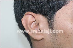
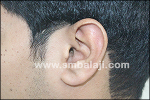
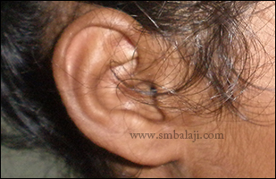
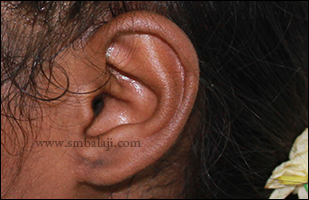
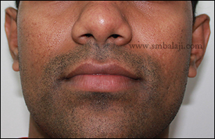
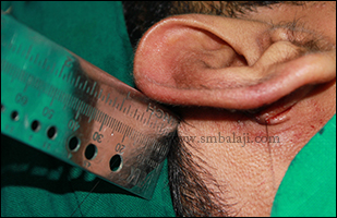
25 year old man reported to our hospital seeking for cosmetic correction of his right ear defect. He consulted many plastic surgeons for the surgical correction but was not satisfied.
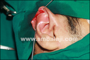
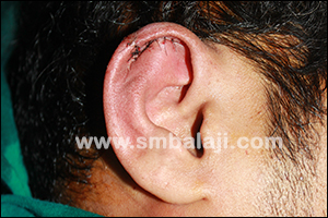
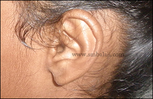
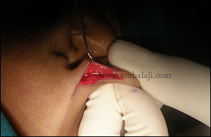
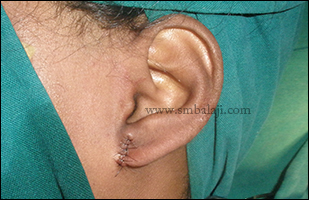
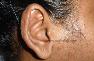
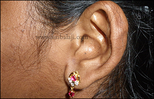
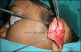
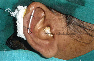
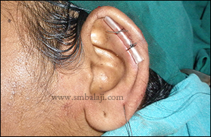
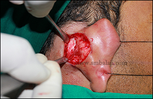
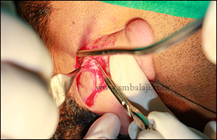
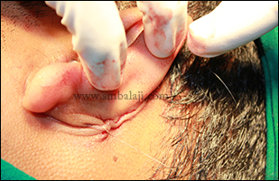
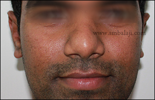
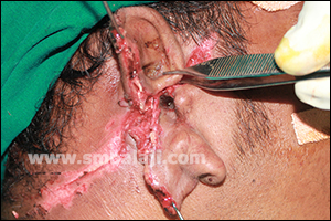
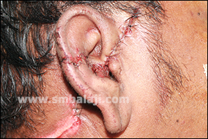
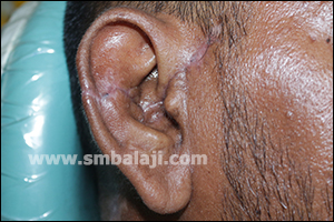
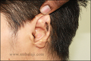
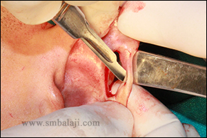
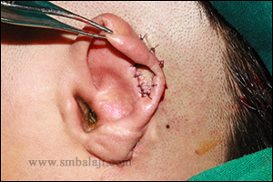
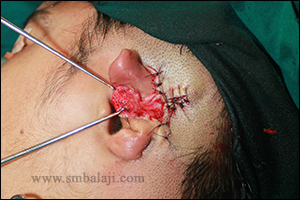
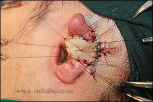
On examination, the helix of the right ear was defective. Maxillofacial Surgeon Dr. S. M. Balaji successfully corrected his ear defect using wedge closure technique without much visible scars. Results are immediate. He is very happy to have single stage ear defect correction.
On examination, the helix of the right ear was defective. Maxillofacial Surgeon Dr. S. M. Balaji successfully corrected his ear defect using wedge closure technique without much visible scars. Results are immediate. He is very happy to have single stage ear defect correction.

 RSS Feed