Best Dental Clinic in Chennai : Best Dental Implant Center in India
FOLLOW US
  • HOME
  • Surgery of The week
  • Clinical Updates
  • Archives
  • Latest News
  • VIDEOS
  • CONTACT US

Surgical removal of the huge ameloblastoma - benign tumor of mandible and reconstruction in a single stage without any scars on the face

11/10/2017

0 Comments

 
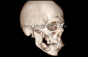
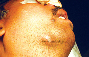
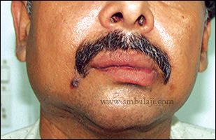
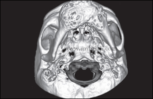
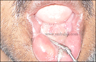
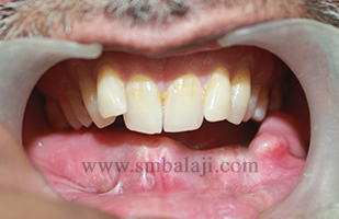
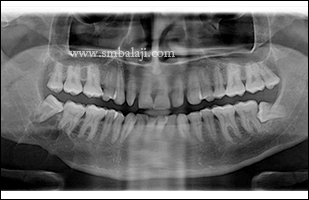
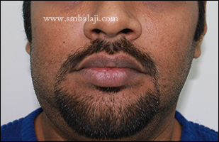
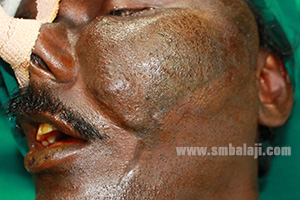
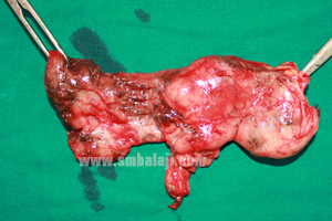
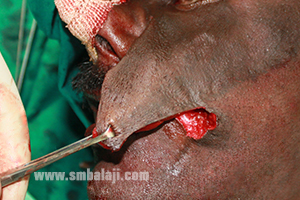
A young man from Singapore reported to me with the complaint of huge swelling in the right side of his face. He added that the size of the swelling was increasing progressively which made his face asymmetrical. He was very cautious about this progressive swelling. Added he had histopathological examination of the intraoral tissue done elsewhere diagnosing as plexiform ameloblastoma.
​
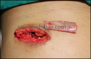
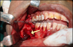
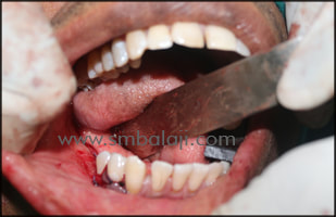
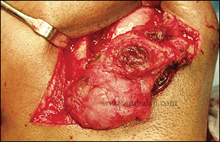
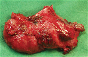
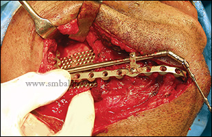
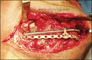
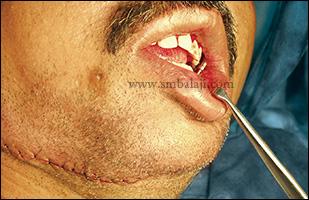
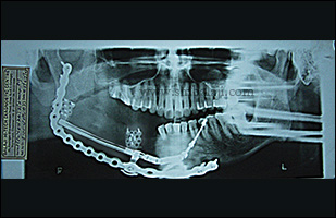
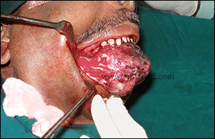
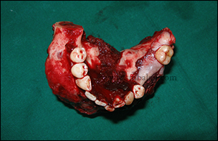
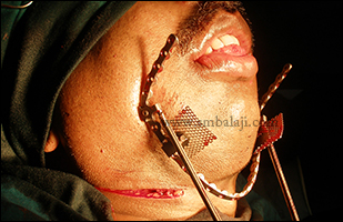
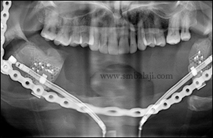
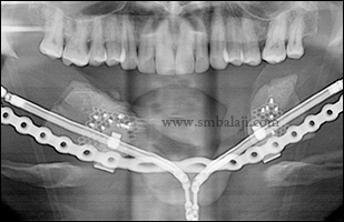
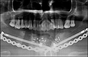
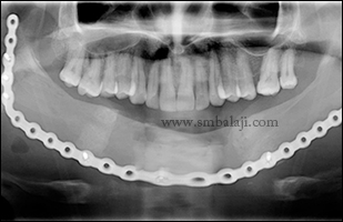
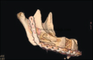
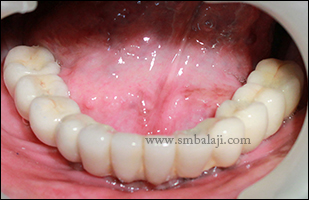
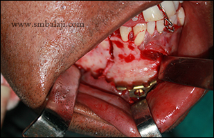
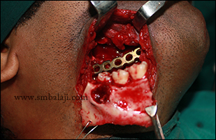
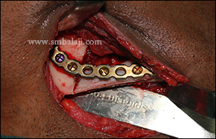
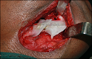
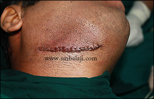
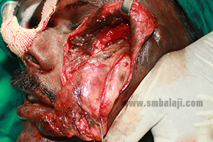
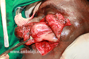
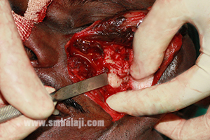
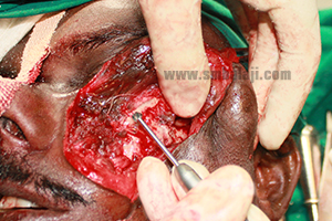
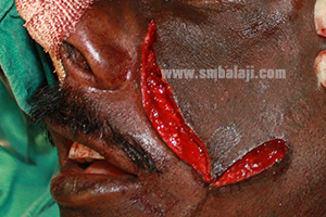
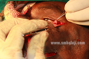
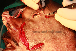
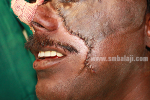
After thorough clinical and radiological examination, Maxillofacial Surgeon  Dr. S. M. Balaji planned to remove the lesion and reconstruct the affected jaw portion in a single stage. Intraorally, complete removal of the benign lesion with sufficient clearance of the bone was done following rib graft which was obtained was fixed to it. Patient was happy to have both removal and reconstruction in a single surgery.
Preoperative 3DCT scan view showing complete labial plate destruction with inferior alveolar canal disruption
Preoperative facial view showing distinguished swelling in right side of face
Rib graft obtained to reconstruct the mandibular defect
Benign tissue removed in toto with involved bone
Intraoral mandibular view after giving enough marginal clearance
Inferior alveolar nerve isolated and preserved
Huge mandibular defect reconstructed using rib graft
Immediately after suturing
0 Comments

Resection of huge mandibular tumor and reconstruction using univector intraoral mandibular distraction technique

16/8/2016

0 Comments

 
This is a case of ameloblastoma involving right side of mandible. Resection and reconstruction of the huge mandibular defect was done without any bone grafting using distraction osteogenesis. Maxillofacial Surgeon Dr. S. M. Balaji successfully corrected using univector intraoral mandibular distraction technique. Simultaneous resection of huge tumor and reconstruction using native bone was done using distraction osteogenesis. By sequential activation of the distractor device, the native bone was made to grow and fill the entire bone defect. Patient feels happy to have both removal and reconstruction of the entire jaw bone defect without any bone graft. 
Preoperative view showing weel defined swelling in the lower border of mandible and note the skin over the swelling is inflammed
Intraoperative view showing exposure of the lesion through submandibular approach followed by resection
Resected specimen
Adaptation and fixation of the plate guided univector unidirectional straight mandibular distractor
Fixation of the mesh foot plates and completion of planned osteotomy
Primary closure of the surgical wound.
Sequential OPG showing the movement of the transport disc with daily activation
Postoperative OPG showing bone formation following distraction osteogenesis
Postoperative view showing maintained contour of the lower jaw without any visible scars on the face
0 Comments

Advanced transport Distraction osteogenesis technique for reconstruction of lower jaw after tumor removal

11/2/2016

0 Comments

 
A 55 year old patient reported to our hospital with a large, bony swelling in the anterior portion of the lower jaw (mandible). He had said that the swelling had gradually increased in size over the past 1 year and there was spontaneous loss of few lower teeth over the last 6 months. He also reported of altered sensation in the lower lip for past 3 weeks which was his main concern.
​
 A 3D CT scan was taken to gauge the extent of the lesion. A biopsy was done which showed the lesion to be a multiple cystic ameloblastoma (tumor) of the lower jaw. Maxillofacial Surgeon Dr. S. M. Balaji removed the entire bony tumor along with the involved bone and reconstructed the defective portion of mandible using the native bone itself without any bone graft by following advanced transport guided distraction osteogenesis technique.

The entire affected portion of the lower jaw was ressected with 1.5 cm marginal clearance followed by reconstruction using titanium plate guided bilateral tripod multi docking complex univector Herford plate guided mandibular transport distractor.

After 5 days of latency period, the distractors were activated at a rate of 0.5mm twice per day (on both sides) until the transport discs had reached the canine region on both sides (amount of distraction on the right side was 46mm and on the left 43.2mm). Following consolidation period of 3 months and radiological evidence of corticalization, the distractors were removed. And complete dental implant rehabilitation done for lower arch in the newly formed healthy bone. Cosmetically well blended fixed ceramic prosthesis given.
X-ray showing tumor in the lower jaw
3DCT scan view showing huge tumor involving more than half of the lower jaw
Intraoperative view showing removal of huge tumor mass intraorally
Complete removal of the tumor mass along with the involved bone in toto
Planning for the fixation of the transport distractor device to the mandible
After tumor removal, fixation of reconstruction plate and segments distracted 1 mm per day
During transport distraction
During distraction
After completion of distraction
After completion of the distraction - facial view
After removal of the distractors and fixing the fragments to the reconstruction plate to achieve the 'U' form of the lower jaw
Healthy new bone formation seen in postoperative 3DCT scan
Postoperative intraoral view showing enhanced healing and augmented new alveolar bone formation
Implants placed in the reconstructed lower jaw
Following dental implant rehabilitation cosmetically well blended fixed ceramic prosthesis placed
0 Comments

Jaw Reconstruction with recombinant technology for tumor resection

29/10/2014

0 Comments

 
A 32 year old man reported to our hospital with the complaint of swelling in the right side of his face. He added that the size of the swelling was increasing progressively which made his face asymmetrical. He was very cautious about this progressive swelling which made him under huge depression.

After thorough clinical and radiological examination, Maxillofacial Surgeon  Dr. S. M. Balaji diagnosed it as ameloblastoma involving the right side angle and ramus of the mandible. Surgery was planned to remove the lesion and reconstruct the affected jaw portion in a single stage. Through submandibular incision, complete removal of the benign lesion along with the involved teeth with sufficient clearance of the bone under General anesthesia.  Long bone plate was fixed along with costochondral graft which was harvested was fixed to it. Miracle protein rhBMP-2 was wrapped over the costochondral graft to induce new bone formation. Incision was closed in layers. Patient was happy to have both removal and reconstruction in a single surgery. Postoperative OPG after 4 months showed good healing with healthy bone formation. 
Preoperative OPG showing huge lesion in the right angle and ramus region of the lower jaw
Preoperative facial view showing swelling in right lower half of the face
Costochondral graft harvested to reconstruct the affected portion of lower jaw
Gingivomucoperiosteal flap raised and affected portion of the lower jaw was exposed after stabilizing with long bone plate
Through submandibula incision wide excision of the benign lesion was done along with the involved teeth in toto
Lingual surface of the excised bone showing complete removal of the affected jaw bone along with the huge benign lesion
Harvested costochondral graft fixed with the bone plate
Harvested graft was wrapped with the miracle protein rhBMP-2 to induce new bone formation
Immediate postoperative view showing complete layer closure of the submandibular incision
4month postoperative OPG showing complete healing of the bone
0 Comments

Successful reconstruction of face after tumor resection

9/4/2014

0 Comments

 
A 40 year old man reported to our hospital with the complaints of huge swelling in the left side of face. He gave the history of previous surgery done elsewhere, were they have removed a tumor extra orally but it got recurred. He also explained about the nature of swelling as he had noticed the swelling 2 years back. But the swelling was small at that time, later it started to grow slowly and extended to the present size now. Initially he had tenderness in the cheek region, but later there was no pain.

After thorough clinical and radiological examinations, it was diagnosed as recurrent Neurofibroma. Maxillofacial surgeon Dr. S. M. Balaji skillfully planned to remove the tumour completely as well as to reconstruct his face. Through the previously operated nasolabial scar, the tumor was exposed by raising the infra orbital skin. Tumor was separated from the maxilla and zygomatic bone and removed completely. Tumor was adherent to the previously operated site which was also detached and removed completely.

The ectropion (turning out of the lower eyelid) in the left eye was repaired with lateral canthotomy incision and orbital septal return flap technique. The inferior tarsal plate was sutured with supra orbital periosteum. Excess skin in the nasolabial area was excised and wound closed in layers.

Pre operative view showing huge swelling in left side of the face extending to lower border of left eye
Incision made in the previously operated naso labial scar region
Tumor mass retrated completely from the adjacent soft tissue
Tumor mass detached from the adjacent soft tissue and in the previously operated scar region
Entire tumor mass removed along with the attachment in the adjacent tissues
Mucosal flap approximation checked
Bony prominence in the tumor region was identified
Bony prominence was reduced with surgical bur
Mucosal flap approximation done
Ectropion in left lower eyelid correction done with lateral canthotomy
Orbital septal return flap technique was done
Enhanced appearance of face with good approximation of soft tissues in layers
0 Comments

    Categories

    All
    Abbe Flap Technique
    Advanced Transport Distraction Osteogenesis
    Advancement Genioplasty
    Alveolar Cleft Repair
    Alveolar Distraction
    Ameloblastoma
    Augmentation Genioplasty
    Augmentation Rhinoplasty
    Bat Ear Correction
    Bilateral Cleft Lip Palate
    Blepharoplasty
    Bone Defect Reconstruction
    Bone Grafting
    Bone Morphogenetic Protein(BMP)
    Broad Flattened Nose Correction
    Bulky Nose Correction
    Chin Correction
    Chin Correction Surgery
    Cleft Lip & Palate Surgery
    Cleft Nose
    Cleft Palate Repair
    Cleft Rhinoplasty
    Complete Facial Rehabilitation
    Complex Facial Fractures
    Condylar Fracture Surgery
    Correction Of Hypoplastic Ramus
    Corrective Jaw Surgery
    Cosmetic Cleft Nose Correction
    Cosmetic Nose Correction
    Cosmetic Rhinoplasty
    Cosmetic Surgery
    Craniofacial Deformities
    Craniofacial Reconstruction
    Craniofacial Surgery
    Cyst Removal
    Dental Implant
    Dentigerous Cyst
    Deviated Nose Correction
    Diplopia
    Distraction Osteogenesis
    Ear Correction
    Ear Defect
    Ear Reconstructive Surgery
    Epiphora
    Extruding Infected MEDPOR
    Eyebrow Correction
    Face Enhancement
    Face Reconstruction
    Facial Asymmetry Correction
    Facial Bone Fracture
    Facial Burn Injury
    Facial Palsy Surgery
    Fibrous Dysplasia
    Flat Nose Correction
    Forehead Bone Fracture
    Forehead Deformity
    Forehead Flap
    Frontal Bossing Correction
    Gummy Smile
    Hemifacial Microsomia
    Hypertelorism Surgery
    Implant
    Incomplete Cleft Lip
    Isolated Cleft Palate Repair
    Jaw Correction Surgery
    Jaw Cyst
    Jaw Deformity
    Jaw Fracture Surgery
    Jaw Joint Ankylosis
    Jaw Reconstructive Surgery
    Jaw Surgery
    Lip Correction
    Lip Defect Correction
    Lip Revision
    Locked Jaw
    Lower Jaw Fracture
    Low Lying Upper Eyelid Correction
    Macrostomia Correction
    Mandible Correction
    Mandible Reconstruction
    Maxillofacial Surgery India
    Maxillomandibular Distraction
    Maxilofacial Surgery
    Micrognathic Mandible Correction
    Microtia
    Nasal Glial Heterotopia
    Naso-orbito-ethmoidal Fracture
    Nose And Lip Correction
    Nose Asymmetry Correction
    Nose Correction
    Nose Job
    Nose Reconstructive Surgery
    Oral Submucous Fibrosis
    Oral Tumor Removal
    Orbital Correction
    Orehead Defect
    Orthognathic Surgery
    Parrot-beak Nose
    Parry Romberg Syndrome
    Pharyngoplasty
    Plastic Surgery
    Primary Cleft Lip Repair
    Profile EnhancemeP
    Reduction Glossectomy
    Reduction Rhinoplasty
    Removal Of Capillary Hemangioma
    RhBMP 2
    RhBMP-2
    Rhinoplasty
    Road Accident Trauma
    Road Accident - Trauma
    Scar Revision
    Secondary Cleft Palate
    Sinus Lift
    Smile Makeover
    Speech Correction
    Square-face Correction
    Surgery Without Scar
    Tessier Nose Defect
    Tissue Expansion
    TMJ Ankylosis
    Tongue Correction
    Tongue Surgery
    Trauma
    Trauma Treatment
    Triangular Shaped Forehead
    Tumor Resection
    Unilateral Cleft Lip
    Unilateral Cleft Lip And Palate
    Unilateral Cleft Lip Repair
    Velopharyngeal Incompetence

    Archives

    April 2021
    March 2021
    January 2021
    December 2020
    April 2020
    March 2019
    February 2019
    January 2018
    November 2017
    October 2017
    September 2017
    June 2017
    May 2017
    April 2017
    March 2017
    January 2017
    December 2016
    October 2016
    September 2016
    August 2016
    July 2016
    June 2016
    May 2016
    April 2016
    March 2016
    February 2016
    January 2016
    December 2015
    October 2015
    September 2015
    August 2015
    July 2015
    June 2015
    May 2015
    April 2015
    March 2015
    February 2015
    January 2015
    December 2014
    November 2014
    October 2014
    September 2014
    August 2014
    July 2014
    June 2014
    May 2014
    April 2014
    March 2014
    February 2014
    January 2014
    December 2013
    November 2013

    RSS Feed

Copyright © 2020 Balaji Dental & Craniofacial Hospital | All rights reserved | Powered by BALAJI DENTAL HOSPITAL