Best Dental Clinic in Chennai : Best Dental Implant Center in India
FOLLOW US
  • HOME
  • Surgery of The week
  • Clinical Updates
  • Archives
  • Latest News
  • VIDEOS
  • CONTACT US

5 year old boy with failed TMJ ankylosis Surgery done elsewhere was corrected successfully by Dr. S. M. Balaji using growth centre transplantation

20/5/2017

0 Comments

 
Left TMJ ankylosis in 5 year old boy showing complete shift of mandible towards the affected side
Preoperative 3DCT showing complete fusion of condylar component to the base of the skull on the left side
Normal TMJ component on right side
Costochondral junction obtained to transfer the growth centre of mandible on left side
Intraoral view showing limited mouth opening
After bony ankylosis release increased mouth opening achieved
Increased mouth opening after few weeks of physiotherapy
Dr S M Balaji examining the dental health and mouth opening of the boy
Dr S M Balaji examining the mouth opening of the boy
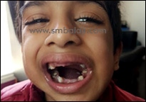
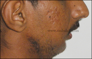
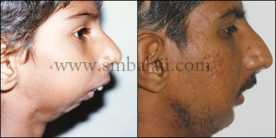
A 5 year old boy was brought to our hospital with a complaint of decreased mouth opening and facial deformity by birth. His parent’s gave the history of multiple surgical correction of TMJ ankylosis done elsewhere. He had unsightly extraoral scar due to previous surgeries. He gave a history of having undergone distraction osteogenesis treatment for the left side of the lower jaw in some other hospital elsewhere, but the treatment subsequently failed.

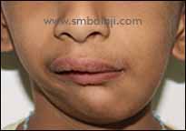
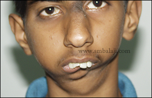
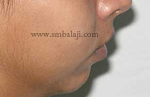
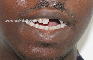
Now on examination he had facial asymmetry, severely retruded lower jaw (mandible), flattened lower face with complete restriction in mouth opening and shift of chin towards left side.
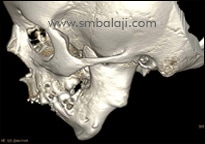
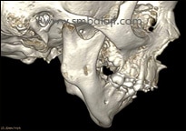
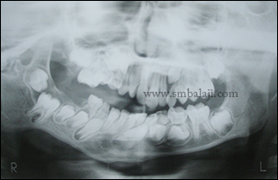
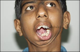
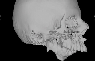
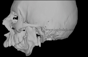
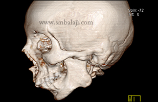
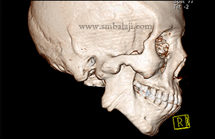
3DCT of facial bones revealed obliterated left temporomandibular (joint jaw) joint space, elongated coronoid process, and reduced ramal height and prominent antegonial notch. The case was diagnosed as true left TMJ bony ankylosis with developing secondary facial deformity. 
​
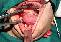
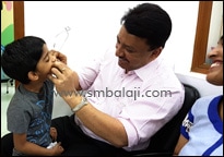
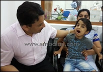
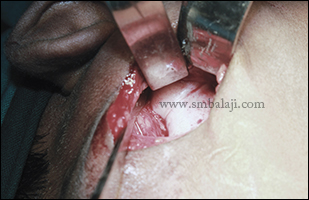
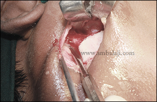
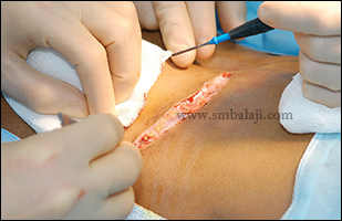
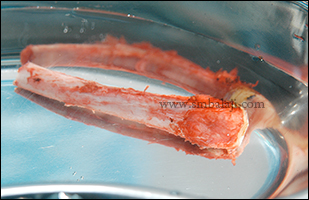
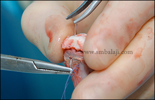
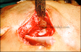
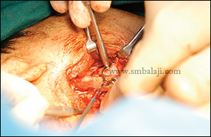
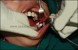
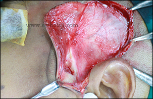
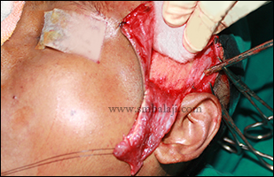
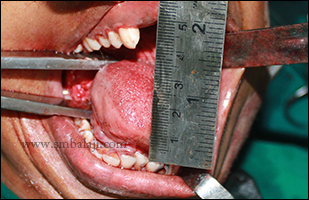
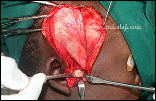
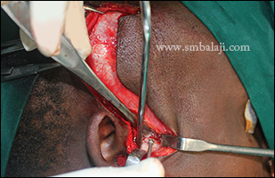
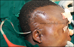
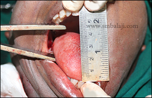
Maxillofacial Surgeon Dr. S. M. Balaji successfully performed bony ankylosis release surgery and transportation of growth centre using costochondral junction obtained from rib cartilage. Postoperative mouth opening was adequate. Following complete dental rehabilitation done to treat the badly broken and decayed teeth, boy’s parents were asked to continue the mouth opening exercise at least 2 months after surgery. 
0 Comments

GROWTH CENTER TRANSPLANTATION USING COSTOCHONDRAL RIB GRAFT FOR BILATERAL TMJ ANKYLOSIS CORRECTION

19/5/2016

0 Comments

 
A 6 year old boy was brought to our hospital with a complaint of decreased mouth opening and facial deformity. No history of trauma elicited by his parents. Extra oral examination revealed severely retruded mandible, flattened lower face and complete restriction in mouth opening.
OPG revealed obliterated right joint space elongated coronoid process, reduced ramal height and prominent antegonial notch. The case was diagnosed as true right TMJ bony ankylosis with developing secondary facial deformity.  
Maxillofacial Surgeon Dr. S. M. Balaji successfully performed gap arthroplasty using temporalis flap interpositioning. Followed by active physiotherapy, mouth opening improved dramatically. But patient failed to be on regular follow up.
After 9 years, exactly at the age of 15, patient reported with the complaint of restricted mouth opening, severe secondary facial deformity and retruded chin giving bird face appearance.
On thorough clinical and radiological examination, it was diagnosed as left TMJ ankylosis with functional right pseudo joint. Dr. S. M. Balaji performed left gap arthroplasty and bilateral condylar component reconstruction using 5th and 7th costochondral rib graft (CCG).
Postoperative mouth opening was adequate. Over 8 years follow up, bilateral CCG was favorable and lower facial projection was improved significantly with increased mouth opening.
Preoperative profile view showing retruded mandible with ill defined chin at the age of 6
Preoperative OPG showing reduced right joint space.
Intraoperative view of the ankylotic mass
Post gap arthroplasty
Preop view at the age of 15 showing severe retrognathic mandible with restricted mouth opening. Note the double chin deformity
Mouth opening with chin deviation and double chin deformity
Profile view showing retruded chin with deficient hyo-mental distance
Preoperative 3DCT view showing hypertrophic coronoid process
Preoperative 3D CT lateral view showing obliterated joint space with hypertrophic coronoid process
Harvesting of the rib graft-following a curvilinear incision layerwise dissection done exposing the right sixth rib
Harvested rib graft with the cartilage cap
Securing of the cartilagenous graft using 4-0 Prolene
Graft introduced into the surgical site for TMJ reconstruction
Securing of the graft at the mandibular posterior border using stainless steel L plates
Postoperative view at the age of 20
Postoperative mouth opening with no chin deviation
Postoperative profile view showing enhanced appearance with prominent chin and increased hyo-mental distance
Pre and postoperative profile view. Note the improvement in facial profile.
0 Comments

Micrognathic mandible correction using simultaneous internal maxillomandibular distraction osteogenesis

17/2/2016

0 Comments

 
22 year old female, a known case of bilateral TMJ ankylosis reported to our hospital for the surgical correction of lower half of the face. Micrognathic mandible present due to retarded development. She gave the history of TMJ ankylosis release surgery done elsewhere during childhood.
After thorough clinical and radiological examination Maxillofacial Surgeon Dr. S. M. Balaji planned to correct the facial asymmetry using internal distraction osteogenesis with maxillomandibular distractor. Bilaterally Paragingival incision was placed over the angle region along the anterior border of ramus. Full thickness mucoperiostal flap was reflected buccally and bone exposed. The direction of the osteotomy cut and positioning of the distractor is the most important as it determines the vector of distraction and this determines the direction of the bone growth. The distractor device was positioned and fixed using screws. Osteotomy is then completed along the medial cortex. Distractor device was checked and wound closure done. Le Fort I osteotomy was completed in maxilla.
After a latency period of 5 days, distraction at the rate of 1 mm per day was accomplished and facial asymmetry was successfully corrected. Thus maxillomandibular distraction technique helped dramatically to enhance the facial appearance without any visible scars and taking autobone graft anywhere from the body. Patient feels very happy to have enhanced facial appearance without any scar on the face.
Retruded mandible due to bilateral temporomandibular joint ankylosis
Retruded mandible with illdefined chin prominence
Intraoperative view showing intraoral fixation of maxillomandibular distractors
Postoperative view after distraction shows forwardly moved mandible without any visible scars on the face
Postoperative profile view showing advanced lower jaw with enhanced chin prominence
0 Comments

Successful correction of TMJ ankylosis with Gap arthroplasty

26/1/2015

0 Comments

 
This was a case of unilateral TMJ ankylosis in a 22 year old girl. She was not able to open her mouth even for taking food for past 20 years. Her upper front teeth were completely decayed and broken. 3DCT scan shows TMJ bony ankylosis on the left side.

Maxillofacial Surgeon Dr. S. M. Balaji planned to open her mouth by following gap arthroplasty with temporalis muscle interpositioning. Under GA, Al-Kayat-Bramley’s incision (Preauricular incision) was made on the left side; a superior limiting cut was made along the demarcation between the rim of the glenoid fossa and the ankylosed head. The ankylotic mass was removed. After ressecting the callous, the gap was further widened by removing adequate bone.

On the left side, the temporalis muscle was rotated and folded inwards into the joint cavity and taken between the medial surface of the ramus taking good care of the adjacent mandibular nerve, facial and maxillary arteries. Then the muscle end was sutured to the medial side of the mandible to provide a submandibular anchorage. A suction drain is placed for 24-36 hrs and the wound was closed in layers. Results are immediate and mouth opening of width up to 44mm was achieved. Patient is put on post surgery physiotherapy to maintain the mouth opening.
Preoperative 3DCT scan showing fused left condyle with that of the cranial base
Inability to open the mouth due to fused condyle on the left side
Preauricular approach to expose the fused joint and cut was made to release the bony fusion
Temporalis muscle interpositioning was done to maintain the movement of the joint
Immediately after relieving the ankylosis mouth opening upto 42mm is achieved
0 Comments

Successful correction of TMJ ankylosis with Gap arthroplasty

21/11/2014

0 Comments

 
This was a case of unilateral TMJ ankylosis in a 24 year old boy from Nigeria. He was not able to open his mouth even for taking food for past 18 years due to an accident. Following the injury, his upper front teeth were lost and he was taking food only through this small gap. 3DCT scan shows TMJ bony ankylosis on the right side and elongated coronoid processes.

Maxillofacial Surgeon Dr. S. M. Balaji planned to open his mouth by following gap arthroplasty with temporalis fascia interpositioning and contralateral coronoidectomy. Under GA, Al-Kayat-Bramley’s incision (Preauricular incision) was made on the right side; a superior limiting cut was made along the demarcation between the rim of the glenoid fossa and the ankylosed head. The ankylotic mass was removed. After ressecting the callous, the gap was further widened by removing adequate bone.

Intraoral coronoidectomy on the contralateral side i.e. the left side was done to facilitate good mouth opening. On the right side, the temporalis muscle was rotated and folded inwards into the joint cavity and taken between the medial surface of the ramus taking good care of the adjacent mandibular nerve, facial and maxillary arteries. Then the muscle end was sutured to the medial side of the mandible to provide a submandibular anchorage. A suction drain is placed for 24-36 hrs and the wound was closed in layers. Results are immediate and mouth opening of inter-incisor width up to 55mm was achieved.
Preoperative view showing negligible mouth opening with missing upper left central and lateral incisors
3DCT scan showing bony fusion of right side condyle with the base of the skull
Preauricular incision in the right side visualizing the ankylosed condyle with that of the glenoid fossa
Exactly in the demarcation between condylar head and glenoid fossa bone cut was made
Temporalis muscle with fascia was detached and interpositioned between condylar head and glenoid fossa and sutured medially in the submandibular region
Immediately after layer closure of the preauricular incision drain out was attached for 24-48 hours to prevent hematoma formation
Immediately after contralateral coronoidectomy mouth opening was increased upto 55mm
0 Comments

    Categories

    All
    Abbe Flap Technique
    Advanced Transport Distraction Osteogenesis
    Advancement Genioplasty
    Alveolar Cleft Repair
    Alveolar Distraction
    Ameloblastoma
    Augmentation Genioplasty
    Augmentation Rhinoplasty
    Bat Ear Correction
    Bilateral Cleft Lip Palate
    Blepharoplasty
    Bone Defect Reconstruction
    Bone Grafting
    Bone Morphogenetic Protein(BMP)
    Broad Flattened Nose Correction
    Bulky Nose Correction
    Chin Correction
    Chin Correction Surgery
    Cleft Lip & Palate Surgery
    Cleft Nose
    Cleft Palate Repair
    Cleft Rhinoplasty
    Complete Facial Rehabilitation
    Complex Facial Fractures
    Condylar Fracture Surgery
    Correction Of Hypoplastic Ramus
    Corrective Jaw Surgery
    Cosmetic Cleft Nose Correction
    Cosmetic Nose Correction
    Cosmetic Rhinoplasty
    Cosmetic Surgery
    Craniofacial Deformities
    Craniofacial Reconstruction
    Craniofacial Surgery
    Cyst Removal
    Dental Implant
    Dentigerous Cyst
    Deviated Nose Correction
    Diplopia
    Distraction Osteogenesis
    Ear Correction
    Ear Defect
    Ear Reconstructive Surgery
    Epiphora
    Extruding Infected MEDPOR
    Eyebrow Correction
    Face Enhancement
    Face Reconstruction
    Facial Asymmetry Correction
    Facial Bone Fracture
    Facial Burn Injury
    Facial Palsy Surgery
    Fibrous Dysplasia
    Flat Nose Correction
    Forehead Bone Fracture
    Forehead Deformity
    Forehead Flap
    Frontal Bossing Correction
    Gummy Smile
    Hemifacial Microsomia
    Hypertelorism Surgery
    Implant
    Incomplete Cleft Lip
    Isolated Cleft Palate Repair
    Jaw Correction Surgery
    Jaw Cyst
    Jaw Deformity
    Jaw Fracture Surgery
    Jaw Joint Ankylosis
    Jaw Reconstructive Surgery
    Jaw Surgery
    Lip Correction
    Lip Defect Correction
    Lip Revision
    Locked Jaw
    Lower Jaw Fracture
    Low Lying Upper Eyelid Correction
    Macrostomia Correction
    Mandible Correction
    Mandible Reconstruction
    Maxillofacial Surgery India
    Maxillomandibular Distraction
    Maxilofacial Surgery
    Micrognathic Mandible Correction
    Microtia
    Nasal Glial Heterotopia
    Naso-orbito-ethmoidal Fracture
    Nose And Lip Correction
    Nose Asymmetry Correction
    Nose Correction
    Nose Job
    Nose Reconstructive Surgery
    Oral Submucous Fibrosis
    Oral Tumor Removal
    Orbital Correction
    Orehead Defect
    Orthognathic Surgery
    Parrot-beak Nose
    Parry Romberg Syndrome
    Pharyngoplasty
    Plastic Surgery
    Primary Cleft Lip Repair
    Profile EnhancemeP
    Reduction Glossectomy
    Reduction Rhinoplasty
    Removal Of Capillary Hemangioma
    RhBMP 2
    RhBMP-2
    Rhinoplasty
    Road Accident Trauma
    Road Accident - Trauma
    Scar Revision
    Secondary Cleft Palate
    Sinus Lift
    Smile Makeover
    Speech Correction
    Square-face Correction
    Surgery Without Scar
    Tessier Nose Defect
    Tissue Expansion
    TMJ Ankylosis
    Tongue Correction
    Tongue Surgery
    Trauma
    Trauma Treatment
    Triangular Shaped Forehead
    Tumor Resection
    Unilateral Cleft Lip
    Unilateral Cleft Lip And Palate
    Unilateral Cleft Lip Repair
    Velopharyngeal Incompetence

    Archives

    April 2021
    March 2021
    January 2021
    December 2020
    April 2020
    March 2019
    February 2019
    January 2018
    November 2017
    October 2017
    September 2017
    June 2017
    May 2017
    April 2017
    March 2017
    January 2017
    December 2016
    October 2016
    September 2016
    August 2016
    July 2016
    June 2016
    May 2016
    April 2016
    March 2016
    February 2016
    January 2016
    December 2015
    October 2015
    September 2015
    August 2015
    July 2015
    June 2015
    May 2015
    April 2015
    March 2015
    February 2015
    January 2015
    December 2014
    November 2014
    October 2014
    September 2014
    August 2014
    July 2014
    June 2014
    May 2014
    April 2014
    March 2014
    February 2014
    January 2014
    December 2013
    November 2013

    RSS Feed

Copyright © 2020 Balaji Dental & Craniofacial Hospital | All rights reserved | Powered by BALAJI DENTAL HOSPITAL