
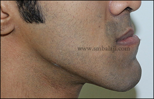
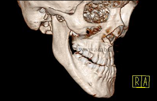
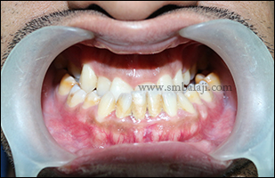
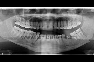
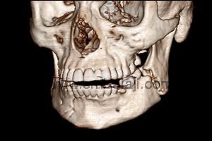
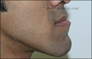
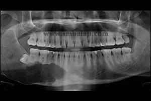
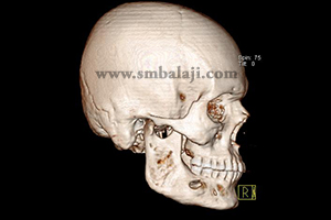
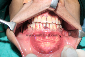
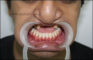
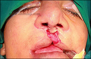
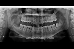
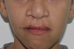
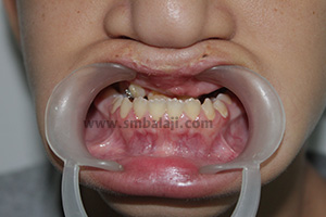
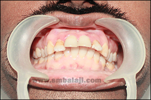
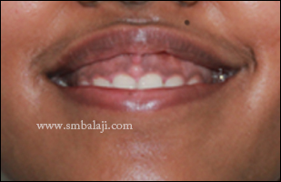
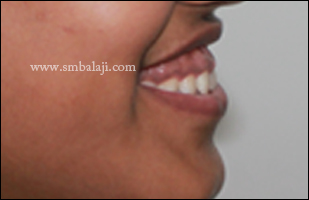
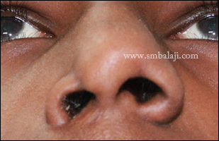
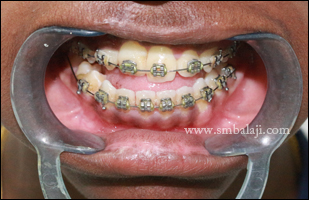
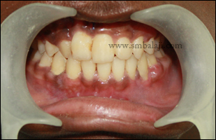
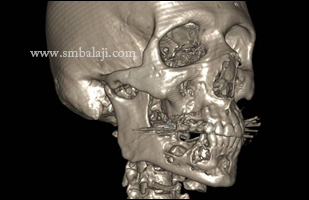
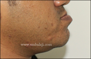
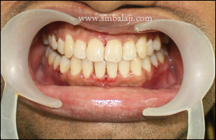
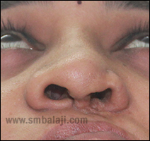
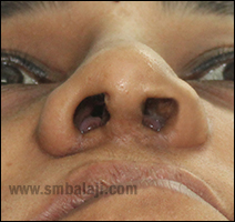
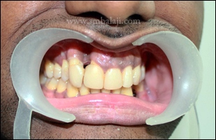
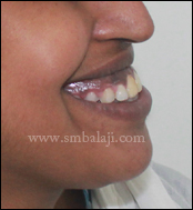
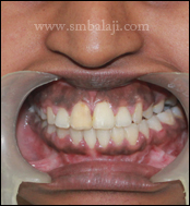
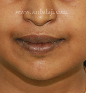
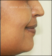
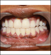
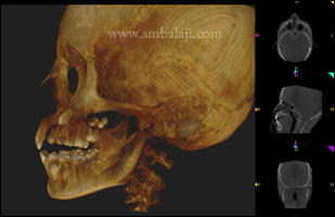
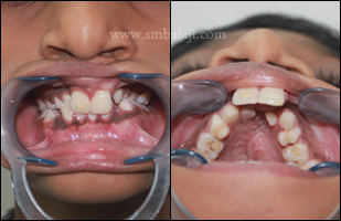
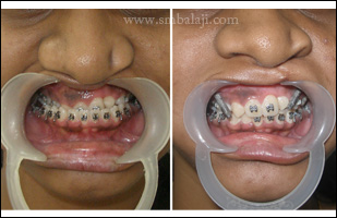
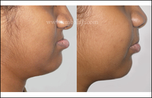
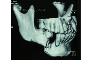
A young man came to our hospital seeking corrective jaw surgery to enhance his appearance. His lower jaw was excessively protruding causing an incorrect teeth occlusion that caused difficulties in biting and chewing. Additionally his chin bone was deviated away from the midline giving his face an asymmetric, crooked appearance.
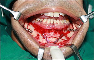
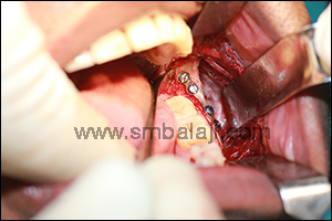
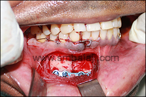
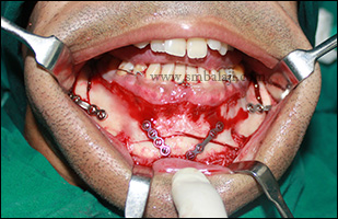
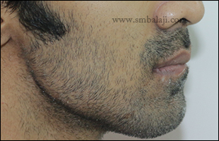
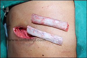
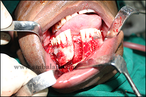
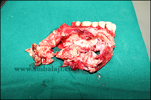
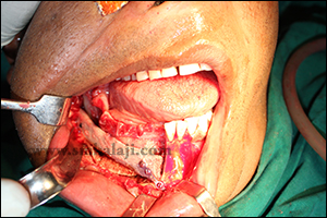
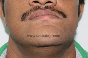
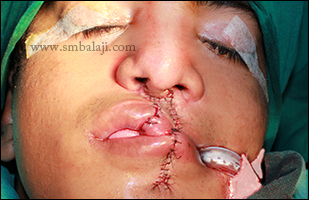
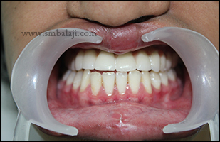
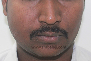
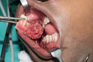
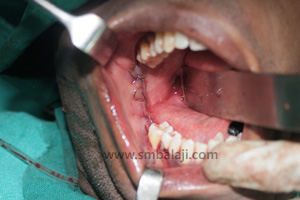
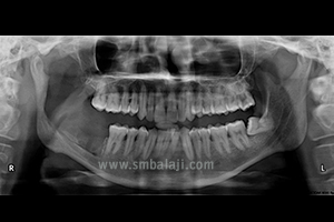
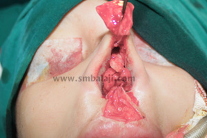
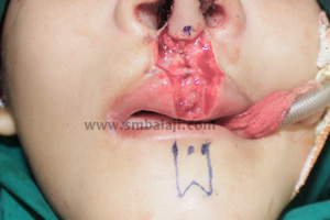
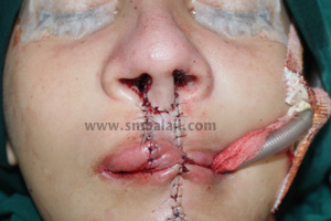
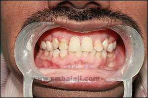
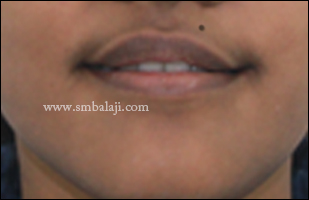
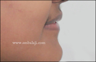
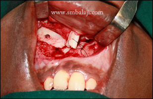
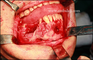
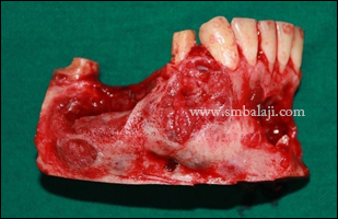
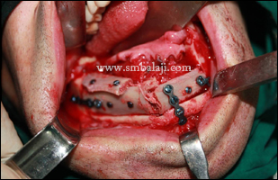
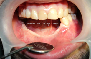
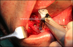
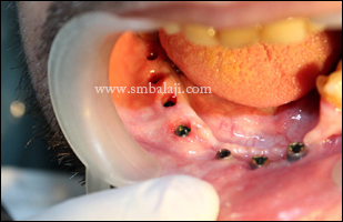
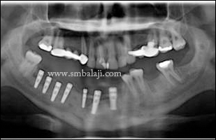
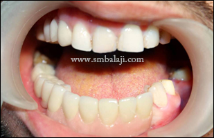
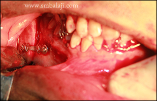
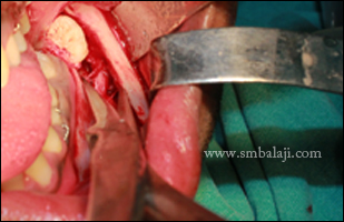
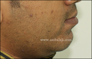
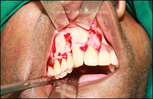
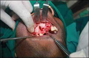
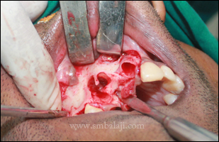
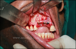
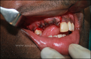
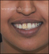
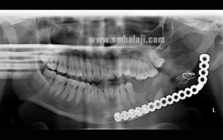
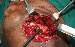
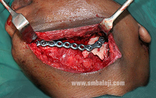
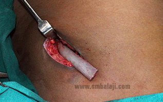
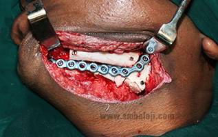
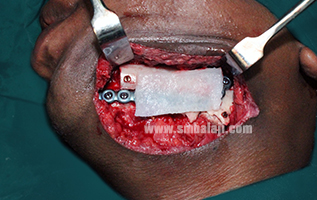
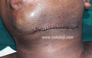
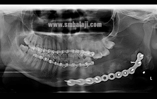
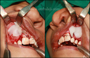
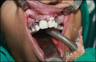
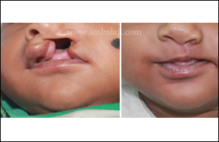
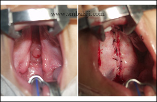
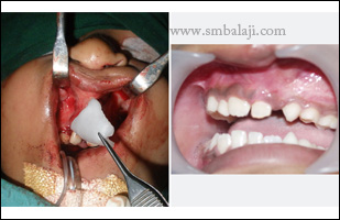
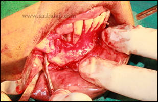
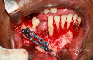
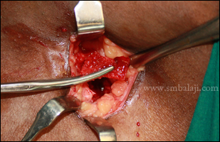
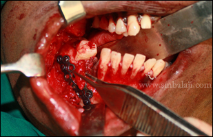
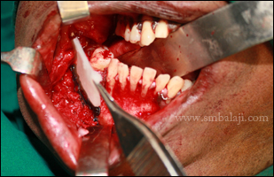
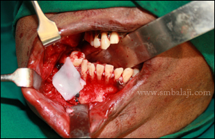
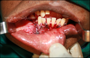
Maxillofacial surgeon Dr. S.M. Balaji planned to correct the jaw defect with a combined bilateral Obwegeser’s sagittal split osteotomy and genioplasty. The mandible was set back to correct the alignment and bite using bilateral Obwegeser’s sagittal split osteotomy. With genioplasty, the deviated chin bone was corrected. The procedure was done from inside the mouth so there were no scars. Immediately after this corrective jaw surgery, the man’s appearance was greatly improved and he is very happy with the enhanced aesthetics.
Maxillofacial surgeon Dr. S.M. Balaji planned to correct the jaw defect with a combined bilateral Obwegeser’s sagittal split osteotomy and genioplasty. The mandible was set back to correct the alignment and bite using bilateral Obwegeser’s sagittal split osteotomy. With genioplasty, the deviated chin bone was corrected. The procedure was done from inside the mouth so there were no scars. Immediately after this corrective jaw surgery, the man’s appearance was greatly improved and he is very happy with the enhanced aesthetics.


 RSS Feed