Best Dental Clinic in Chennai : Best Dental Implant Center in India
FOLLOW US
  • HOME
  • Surgery of The week
  • Clinical Updates
  • Archives
  • Latest News
  • VIDEOS
  • CONTACT US

Craniofacial Reconstruction of 5-Year-Old Girl has done By  Dr. S. M. Balaji.

8/12/2016

0 Comments

 
Preoperative photograph of Jasra. Please note the facial asymmetry.
Customized 3-D stereolithographic models of Jasra’s skull before surgery, frontal view
Customized 3-D stereolithographic models of Jasra’s skull before surgery, left lateral view
Dr. S. M. Balaji operating on Jasra
Mock surgery performed on the customized 3-D stereolithographic models of Jasra’s skull before the actual surgery, frontal view
Mock surgery performed on the customized 3-D stereolithographic models of Jasra’s skull before the actual surgery, left lateral view
An intraoperative photograph of the sonic weld resorbable plates used to fix the medially rotated frontal bone advancement of Jasra’s skull
Jasra, two weeks postoperative, left lateral view
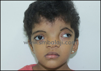
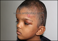
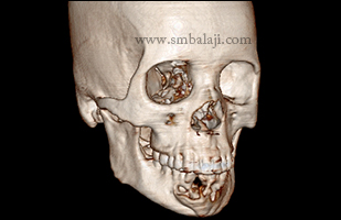
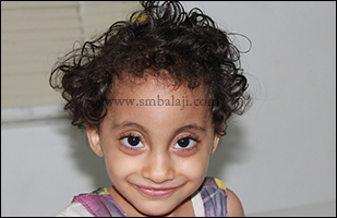
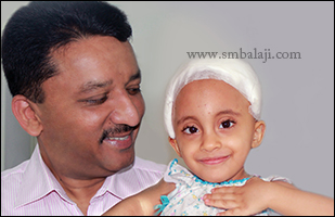
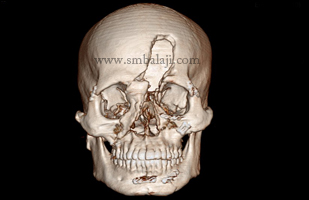
Ms. Jasra is a 5 year old girl who presented to Dr. S. M. Balaji with craniosynostosis with left plagiocephaly.  This had resulted in her having a deformed head.  Plagiocephaly by definition is flattening of one part of the skull, which leads to facial deformity. She had previously undergone cranial/frontal bone reconstruction and orbital rim advancement elsewhere.
​
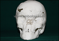
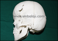
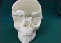
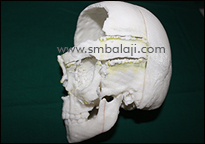
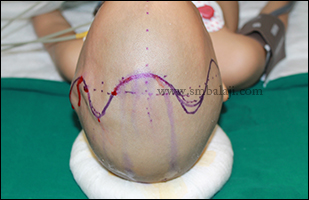
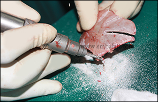
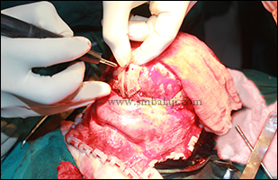
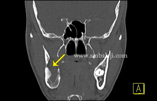
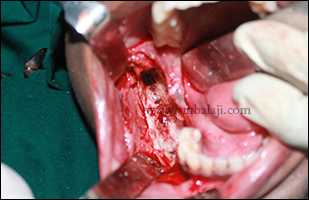
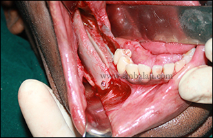
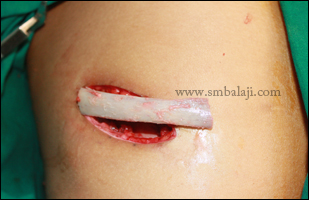
Craniofacial Surgeon Dr. S. M. Balaji decided to proceed with a craniostomy with calvarial reconstruction to correct her deformity. Customized 3-D stereolithographic models were used for presurgical planning.

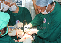
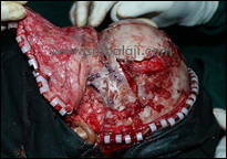
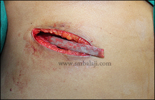
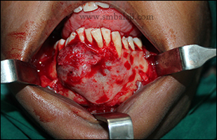
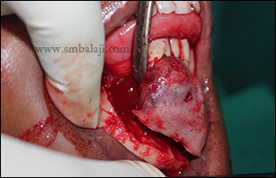
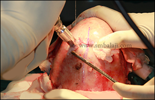
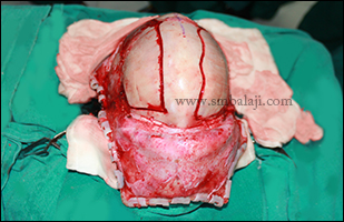
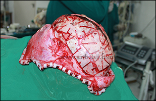
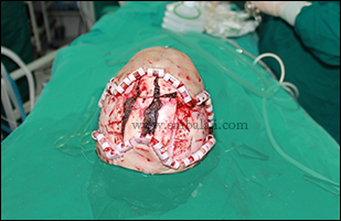
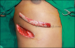
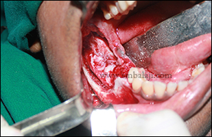
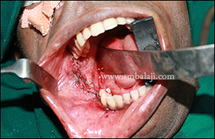
She underwent the cranial vault reconstruction surgery on October 24, 2016, at Balaji Dental and Craniofacial Hospital, Chennai by Dr. S. M. Balaji as part of the preconference live surgical demonstration prior to the 10th World Cleft Lip, Palate, and Craniofacial Congress.  Dr. Balaji used sonic weld bioresorbable plates for rotation and advancement of the frontal bone complex. This is an essential surgery because not doing this surgery would result in compromise of brain development, which would lead to decreased mental acuity, vision problems, and many other cranial functions.  Sonic weld plates were used instead of titanium plates because they resorb over time thus facilitating normal bone growth and brain development as opposed to the permanent titanium plates, which would require another surgery for their removal.

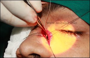
The parents expressed their appreciation of how Dr. S. M. Balaji was helping in transforming Jasra’s life for the better. Jasra would need facial asymmetry corrective surgeries at a later date.
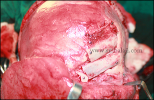
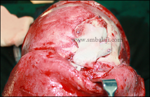
0 Comments

Successful surgical removal and reconstruction of the ameloblastoma of mandible

13/4/2016

0 Comments

 
A 26 year old woman reported to our hospital with the complaint of swelling in the right side of her face. She added that the swelling was increasing in size dramatically and the lower anterior teeth are mobile teeth. She was very cautious about this progressive swelling which made her under huge depression.
​
After thorough clinical and radiological examination, Maxillofacial Surgeon Dr. S. M. Balaji diagnosed ameloblastoma extending from right side of the body of the mandible till the other side. Surgery was planned to remove the lesion and reconstruct the affected jaw portion in a single stage. Through gingivomucoperiosteal flap, complete removal of the lesion along with the involved teeth with sufficient clearance of the bone was done under General anesthesia.  Costochondral rib graft was harvested and fixed to it. Flap was approximated and sutured. Patient was happy to have both removal and reconstruction in a single surgery.
Preoperative OPG showing lesion in the right body of mandible extending to the left side premolar region
Preoperative 3DCT view showing huge osteolytic lesion involving the labial and lingual cortex of anterior region of mandible
Intraoral view showing huge swelling and cortical expansion in anterior region of mandible
Costochondral graft harvested to reconstruct the affected portion of lower jaw
Intraoperative view showing huge bony tumour involving the entire anterior portion of mandible
Gingivomucoperiosteal flap raised and affected portion of the lower jaw was exposed
Resected portion of mandible with the tumour and involved teeth in toto
Wide excision of the benign lesion was done along with the involved teeth in toto
Harvested costochondral graft fixed with the adjacent bone using bone plates
Immediately after suturing
0 Comments

Total Calvarial Reconstruction for Metopic cranial synostosis (Trigonocephaly)

1/9/2015

0 Comments

 
Preoperative view
Note the triangular forehead deformity
Marking for the bicoronal incision
Craniotomy using Advanced, self-stopping, rotatory craniotome
Intraoperative view showing the manner in which bone has been cut
Dissected bone fragment with multiple releasing cuts for future growth
Dissected specimen for reassembly
Fixing of bone fragments using resorbable plates and screws using special screw driver
Assembly of bone fragment
Reapproximated bone fragments with proper positioning
Reorientation of entire skull cap
Post operative view
Picture
Post operative view
A 3 year old girl from Iraq reported with triangular shaped forehead deformity. Patient was evaluated and diagnosed as a case of nonsyndromic trigonocephaly, which is a premature fusion of the bone joint along the middle of the forehead called the metopic suture. As the early fusion of bone does not permit the growth of the brain it will lead to the increased pressure causing cognitive impairment & delayed mile stones. Owing to these dangerous effects with the progressive growth, her parents brought her to our hospital. Maxillofacial Surgeon Dr. S.M. Balaji successfully performed Craniotomy with Total Calvarial Reconstruction with the goal of aligning the midsutural fused area thereby preventing all associated complication.

Using an advanced, self-stopping, rotatory craniotome, the cranium is cut in to several pieces. The entire skull crown is dissected in several pieces and reassembled to accommodate the growth of the brain with suitable repositioning. Then the re-approximated bones are held together by a bioresorbable plate and screws. The specially designed screw driver is used which creates a small electric discharge creating a strong anchorage holding the bone in place. The entire skull cap is reoriented, approximated and fixed. The change in skull is visible in the immediate post-operative period. The usage of bioresorbable screw and plate removes the requirement of a re-surgery to remove the plates.

In future, the brain would be accommodated in the enlarged, spacious cranial vault thus all the dangerous complications have been evaded.
0 Comments

Successful reconstruction of infected portion of mandible due to sequestrum of failed bovine bone grafting done elsewhere

2/5/2015

0 Comments

 
A 30 year old man came to our hospital with the complaints of swelling in right side of face with pain for past 3 months. He gave the history that he had a huge osteolytic lesion involving right side of the mandible which was removed and reconstructed with bovine bone grafting elsewhere. 

Due to graft rejection there was a huge infected cyst with sequestrum (Dead bone) in the involved region of mandible. Intraorally there was an incomplete closure of buccal mucosa where the sequestrum was exposed. After thorough clinical and radiological examination, Maxillofacial Surgeon Dr. S. M. Balaji successfully removed the dead bone with the infected cyst intraorally and reconstructed the same with costochondral rib graft. Patient feels happy to have both the removal and reconstruction of the infected portion of mandible without any visible surgical marks on the face.
Preoperative 3DCT scan view showing complete destruction of ramus of mandible on right side with infected cyst
Harvested costochondral rib graft used to reconstruct the defective portion of mandible
Infected portion of mandible surgically exposed showing sequestrum of the failed bovine bone graft done elsewhere
After complete removal of the cyst along with sequestrum of the bovine bone graft
Harvested rib graft used to reconstruct the defective portion of mandible
Immediately after suturing
0 Comments

Complex forehead & cheek bone fracture reconstruction with abnormal tear flow correction

20/6/2014

0 Comments

 
A 27-year-old man from Kolkata reported to our hospital seeking expert craniofacial reconstruction. He had met with a vehicular accident few months back and suffered fracture of the forehead, cheek bones & lower jaw. He underwent initial surgery elsewhere. He had a gross skull defect with a depression in his left forehead. He also complained of continuous tears running down his face from his left eye.

3D CT scan revealed fracture of the frontal bone on the left side. Due to the traumatic injury, a defect in the nasolacrimal duct caused epiphora or abnormal tear flow. The normal flow of tears from the eye to the nose was damaged.

Craniofacial surgeon Dr. S.M. Balaji skillfully performed the complex reconstruction. A cortical bone graft was harvested from the rib and used to reconstruct the left forehead. Miracle protein rhBMP-2 was placed on the bone graft to stimulate new bone formation.

The defect in the nasolacrimal duct was corrected with DCR or Dacryocystorhinostomy. The ostium was opened with rongeurs, bone adjacent to nasolacrimal sac was removed. A new direct communication was created between the nasolacrimal sac & the nasal mucosa by placing a silicone tube to enable the tears to drain into the nose. Complete craniofacial defect was corrected successfully.
Preoperative 3DCT scan view showing fractured frontal bone involving supraorbital rim with commuited fracture of nasal bone on the left side
Cortical graft harvested from the ribs for frontal bone reconstruction
Bicoronal flap raised and costal grafts were fixed with the frontal process with mini bone screws and stabilised
Bone graft was covered with miracle protein rhBMP-2 to induce new bone formation
DCR was performed by creating new communication between nasolacrimal sac and nasal mucosa with silicone tube
0 Comments

    Categories

    All
    Abbe Flap Technique
    Advanced Transport Distraction Osteogenesis
    Advancement Genioplasty
    Alveolar Cleft Repair
    Alveolar Distraction
    Ameloblastoma
    Augmentation Genioplasty
    Augmentation Rhinoplasty
    Bat Ear Correction
    Bilateral Cleft Lip Palate
    Blepharoplasty
    Bone Defect Reconstruction
    Bone Grafting
    Bone Morphogenetic Protein(BMP)
    Broad Flattened Nose Correction
    Bulky Nose Correction
    Chin Correction
    Chin Correction Surgery
    Cleft Lip & Palate Surgery
    Cleft Nose
    Cleft Palate Repair
    Cleft Rhinoplasty
    Complete Facial Rehabilitation
    Complex Facial Fractures
    Condylar Fracture Surgery
    Correction Of Hypoplastic Ramus
    Corrective Jaw Surgery
    Cosmetic Cleft Nose Correction
    Cosmetic Nose Correction
    Cosmetic Rhinoplasty
    Cosmetic Surgery
    Craniofacial Deformities
    Craniofacial Reconstruction
    Craniofacial Surgery
    Cyst Removal
    Dental Implant
    Dentigerous Cyst
    Deviated Nose Correction
    Diplopia
    Distraction Osteogenesis
    Ear Correction
    Ear Defect
    Ear Reconstructive Surgery
    Epiphora
    Extruding Infected MEDPOR
    Eyebrow Correction
    Face Enhancement
    Face Reconstruction
    Facial Asymmetry Correction
    Facial Bone Fracture
    Facial Burn Injury
    Facial Palsy Surgery
    Fibrous Dysplasia
    Flat Nose Correction
    Forehead Bone Fracture
    Forehead Deformity
    Forehead Flap
    Frontal Bossing Correction
    Gummy Smile
    Hemifacial Microsomia
    Hypertelorism Surgery
    Implant
    Incomplete Cleft Lip
    Isolated Cleft Palate Repair
    Jaw Correction Surgery
    Jaw Cyst
    Jaw Deformity
    Jaw Fracture Surgery
    Jaw Joint Ankylosis
    Jaw Reconstructive Surgery
    Jaw Surgery
    Lip Correction
    Lip Defect Correction
    Lip Revision
    Locked Jaw
    Lower Jaw Fracture
    Low Lying Upper Eyelid Correction
    Macrostomia Correction
    Mandible Correction
    Mandible Reconstruction
    Maxillofacial Surgery India
    Maxillomandibular Distraction
    Maxilofacial Surgery
    Micrognathic Mandible Correction
    Microtia
    Nasal Glial Heterotopia
    Naso-orbito-ethmoidal Fracture
    Nose And Lip Correction
    Nose Asymmetry Correction
    Nose Correction
    Nose Job
    Nose Reconstructive Surgery
    Oral Submucous Fibrosis
    Oral Tumor Removal
    Orbital Correction
    Orehead Defect
    Orthognathic Surgery
    Parrot-beak Nose
    Parry Romberg Syndrome
    Pharyngoplasty
    Plastic Surgery
    Primary Cleft Lip Repair
    Profile EnhancemeP
    Reduction Glossectomy
    Reduction Rhinoplasty
    Removal Of Capillary Hemangioma
    RhBMP 2
    RhBMP-2
    Rhinoplasty
    Road Accident Trauma
    Road Accident - Trauma
    Scar Revision
    Secondary Cleft Palate
    Sinus Lift
    Smile Makeover
    Speech Correction
    Square-face Correction
    Surgery Without Scar
    Tessier Nose Defect
    Tissue Expansion
    TMJ Ankylosis
    Tongue Correction
    Tongue Surgery
    Trauma
    Trauma Treatment
    Triangular Shaped Forehead
    Tumor Resection
    Unilateral Cleft Lip
    Unilateral Cleft Lip And Palate
    Unilateral Cleft Lip Repair
    Velopharyngeal Incompetence

    Archives

    April 2021
    March 2021
    January 2021
    December 2020
    April 2020
    March 2019
    February 2019
    January 2018
    November 2017
    October 2017
    September 2017
    June 2017
    May 2017
    April 2017
    March 2017
    January 2017
    December 2016
    October 2016
    September 2016
    August 2016
    July 2016
    June 2016
    May 2016
    April 2016
    March 2016
    February 2016
    January 2016
    December 2015
    October 2015
    September 2015
    August 2015
    July 2015
    June 2015
    May 2015
    April 2015
    March 2015
    February 2015
    January 2015
    December 2014
    November 2014
    October 2014
    September 2014
    August 2014
    July 2014
    June 2014
    May 2014
    April 2014
    March 2014
    February 2014
    January 2014
    December 2013
    November 2013

    RSS Feed

Copyright © 2020 Balaji Dental & Craniofacial Hospital | All rights reserved | Powered by BALAJI DENTAL HOSPITAL