
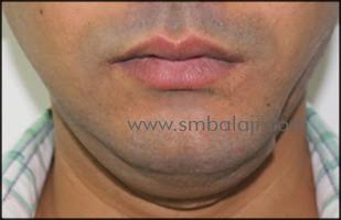
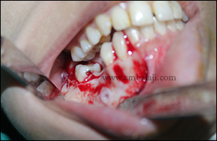
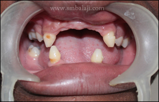
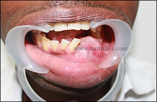
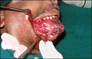
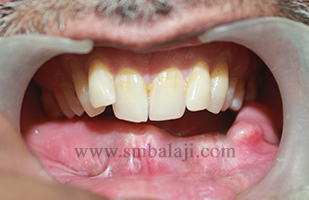
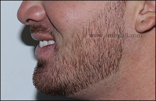
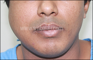
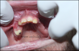
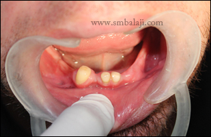
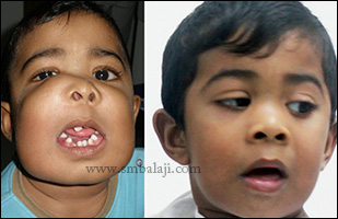
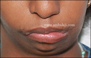
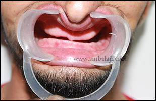
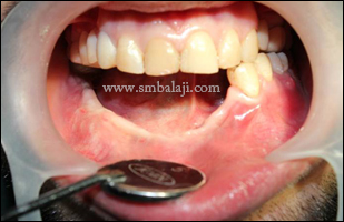
A 30-year-old man from Bangladesh was suffering from severe pain and swelling in his right side of lower jaw. He also complained of difficulty to open his mouth wide and bite and chew foods. He was advised by his family doctor in his home town to go to Balaji Dental and Craniofacial Hospital at Chennai, India, for specialized treatment.
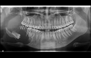
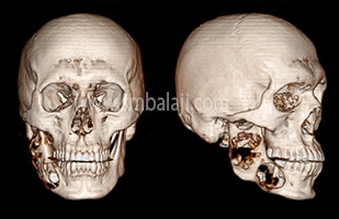
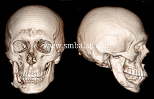
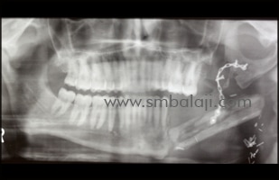
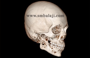
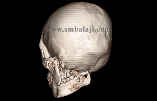
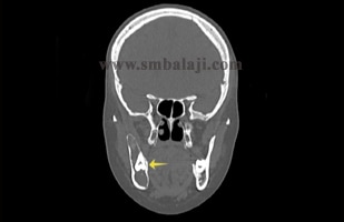
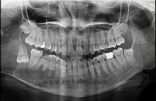
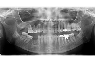
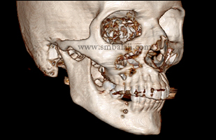
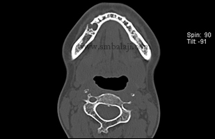
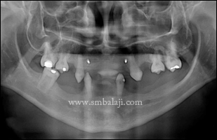
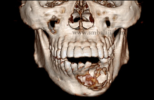
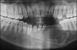
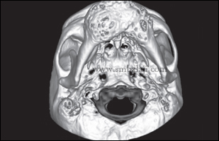
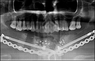
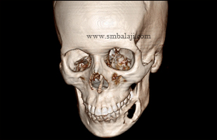
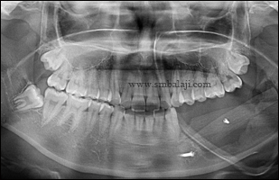
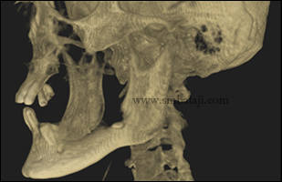
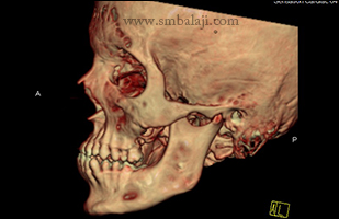
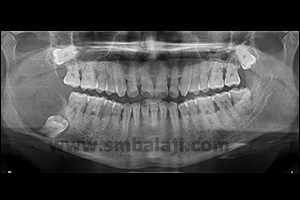
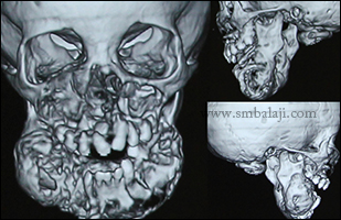
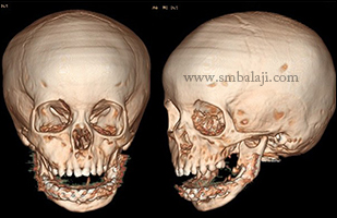
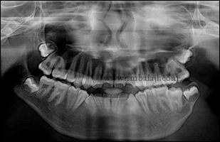
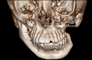
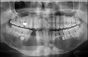
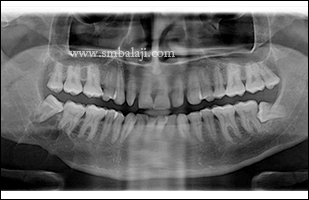
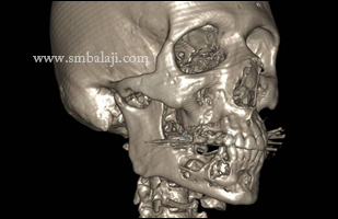
Maxillofacial Surgeon Dr. S.M. Balaji thoroughly evaluated his jaw defect clinically and using advanced 3D CT scan. The scan images showed that he had multiple and extensive areas of bone loss that appeared to resemble large cystic tumors involving the almost entire right side of lower jaw.
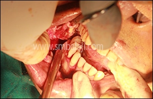
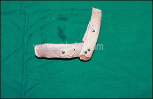
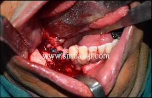
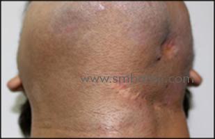
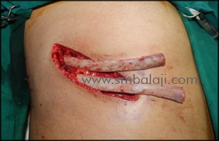
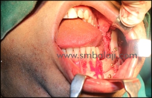
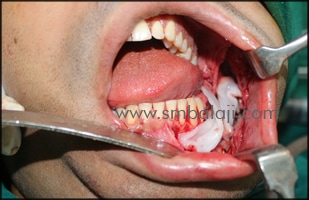
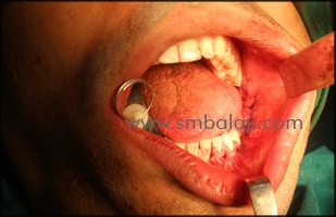
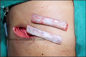
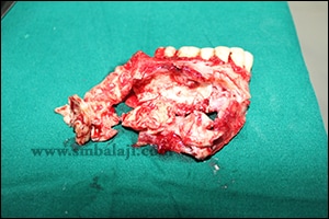
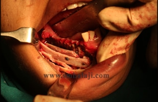
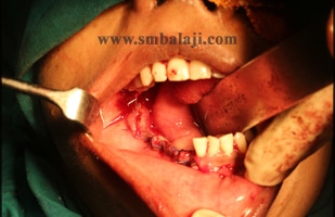
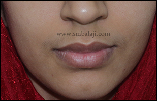
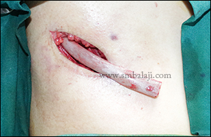
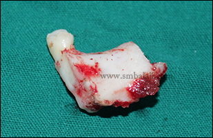
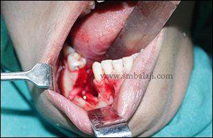
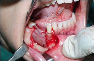
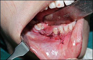
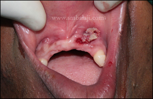
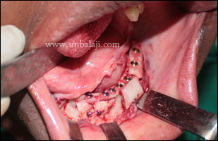
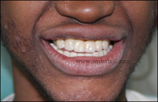
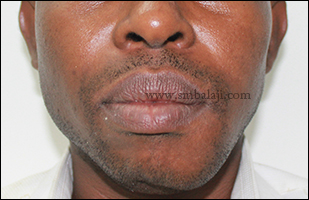
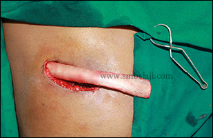
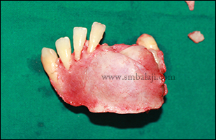
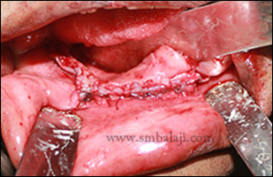
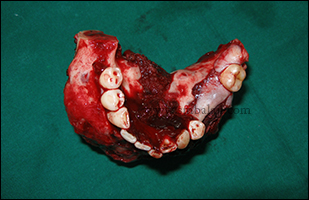
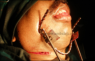
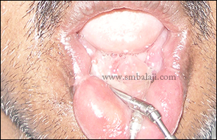
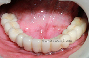
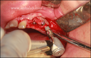
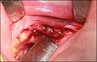
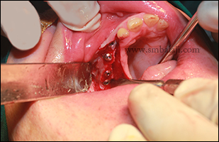
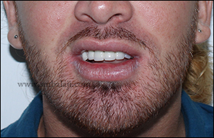
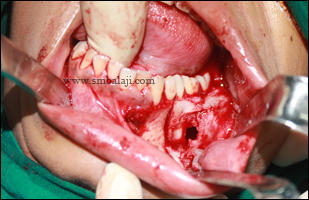
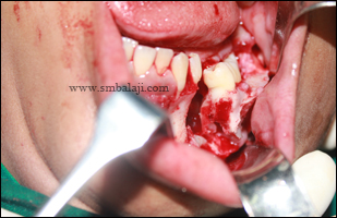
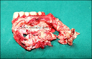
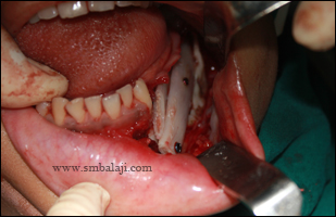
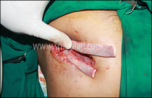
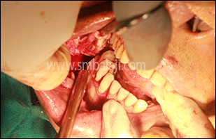
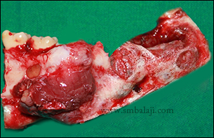
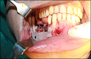
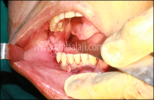
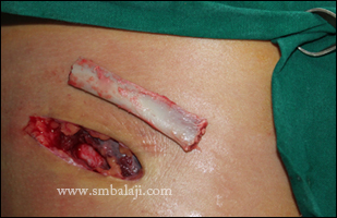
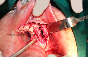
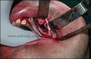
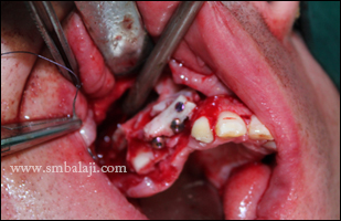
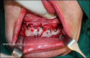
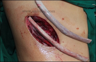
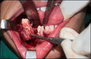
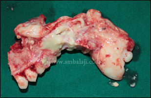
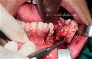
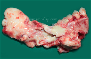
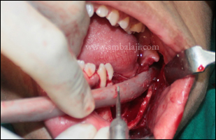
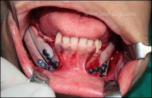
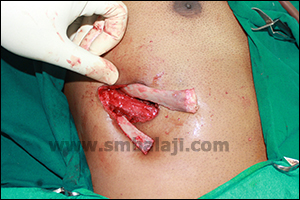
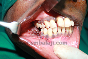
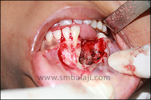
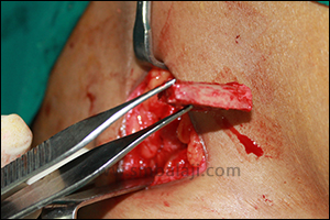
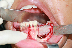
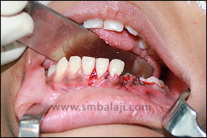
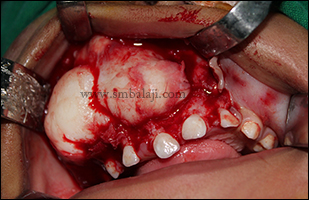
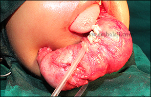
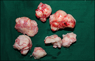
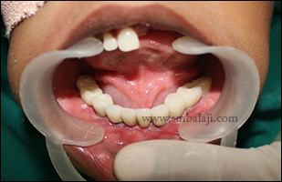
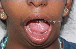
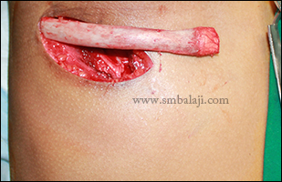
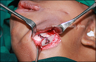
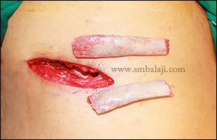
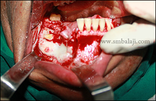
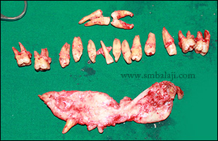
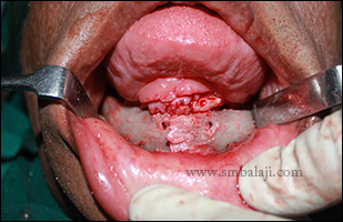
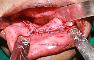
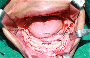
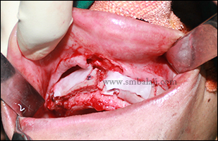
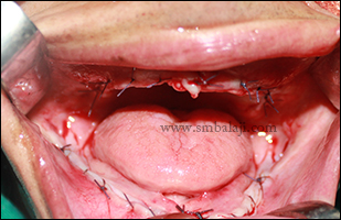
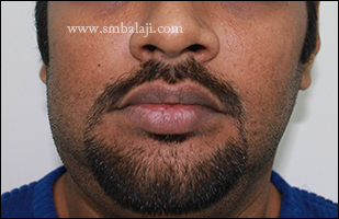
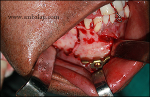
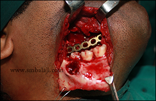
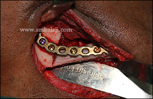
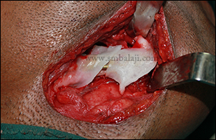
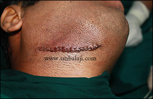
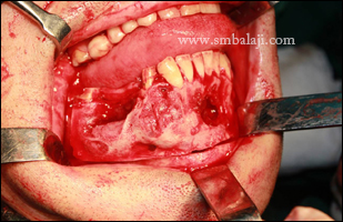
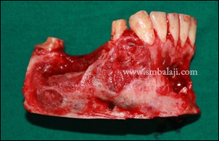
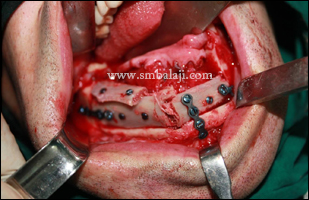
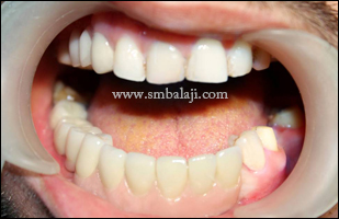
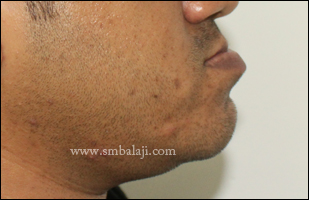
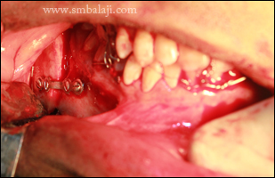
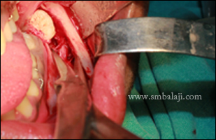
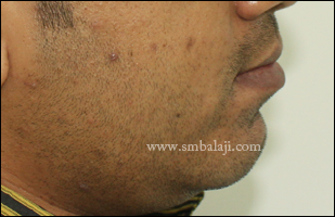
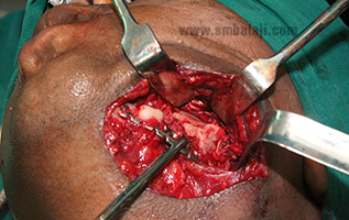
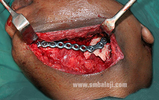
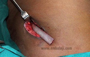
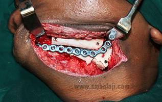
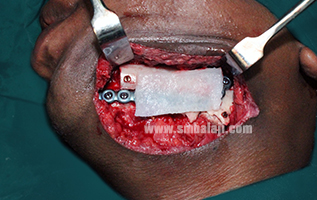
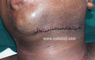
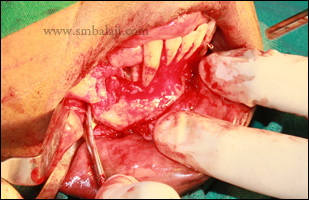
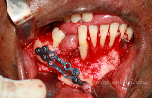
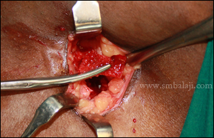
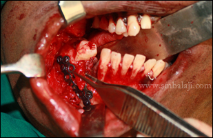
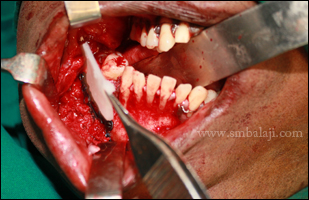
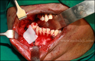
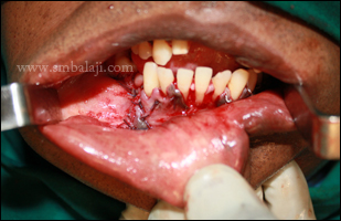
Biopsy of the lesion was done which proved it to be odontogenic keratocysts. Dr. Balaji planned a complete reconstruction of the lower jaw right side, using the man’s own rib graft. The affected portion of the lower jaw bone was surgically removed. Diseased portions of the bone were completely and thoroughly removed to ensure that there is no recurrence. A costochondral cartilage graft (rib graft) was taken and used to reconstruct the lower law. The man feels very happy for the surgery outcome and the entire surgery was done intra orally avoiding external scar formation.
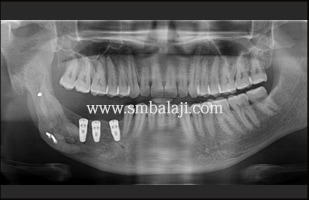
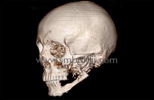
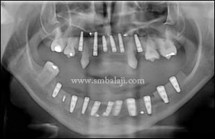
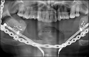
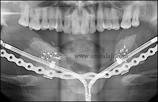
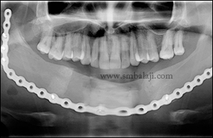
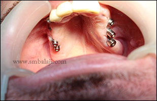
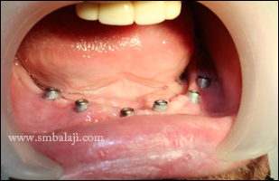
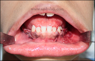
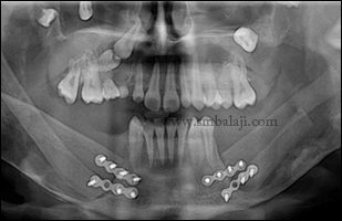
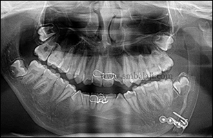
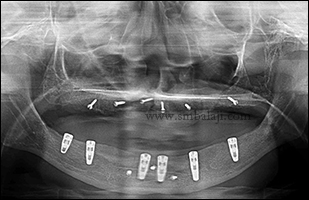
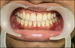
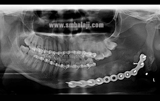
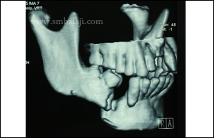
4 months follow-up
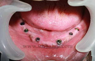
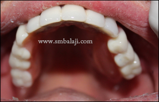
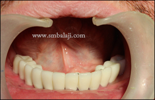
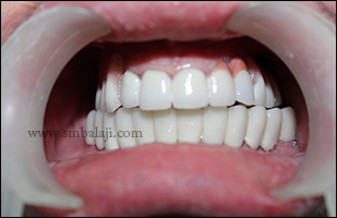
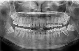
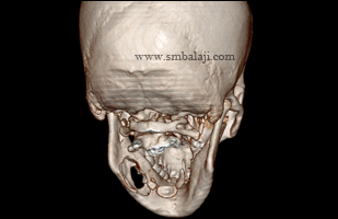
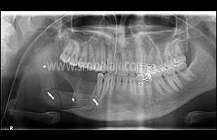
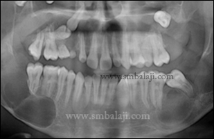
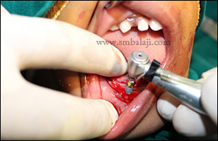
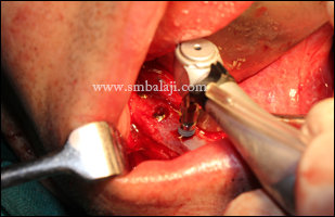
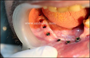
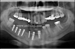
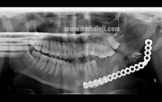
4 months postoperative digital OPG and 3DCT shows good bone formation. Dental implants fixed in the newly formed bone with good stability and retention.
Maxillofacial Surgeon Dr. S.M. Balaji thoroughly evaluated his jaw defect clinically and using advanced 3D CT scan. The scan images showed that he had multiple and extensive areas of bone loss that appeared to resemble large cystic tumors involving the almost entire right side of lower jaw.
Biopsy of the lesion was done which proved it to be odontogenic keratocysts. Dr. Balaji planned a complete reconstruction of the lower jaw right side, using the man’s own rib graft. The affected portion of the lower jaw bone was surgically removed. Diseased portions of the bone were completely and thoroughly removed to ensure that there is no recurrence. A costochondral cartilage graft (rib graft) was taken and used to reconstruct the lower law. The man feels very happy for the surgery outcome and the entire surgery was done intra orally avoiding external scar formation.
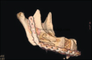
4 months follow-up
4 months postoperative digital OPG and 3DCT shows good bone formation. Dental implants fixed in the newly formed bone with good stability and retention.


 RSS Feed