Best Dental Clinic in Chennai : Best Dental Implant Center in India
FOLLOW US
  • HOME
  • Surgery of The week
  • Clinical Updates
  • Archives
  • Latest News
  • VIDEOS
  • CONTACT US

Successful correction of jaw joint ankylosis (locked jaw) to increase mouth-opening

28/5/2015

0 Comments

 
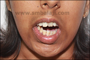
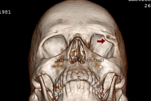
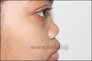
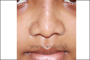
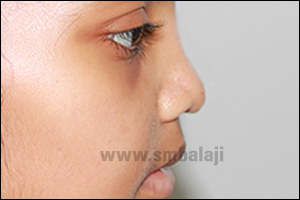
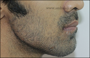
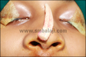
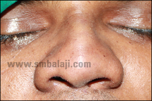
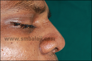
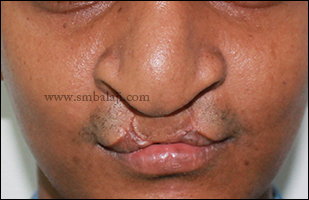
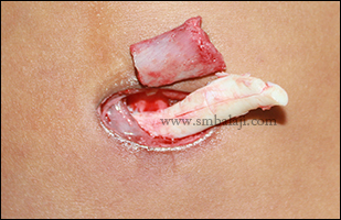
An 18-year-old girl was brought to our hospital by her parents for expert treatment of her jaw joint defect. The girl had severely restricted mouth opening and her lower jaw deviated to one side when she opened her mouth. She had great difficulty in eating and speaking. She gave a history of a fall injury to her chin in her childhood.

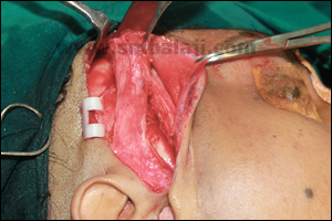
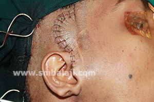
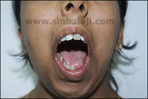
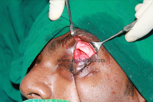
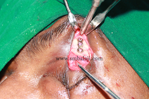
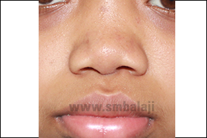
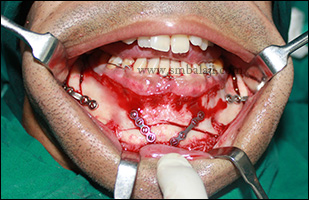
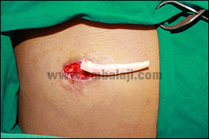
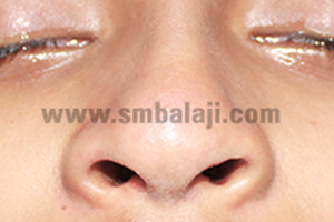
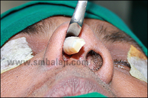
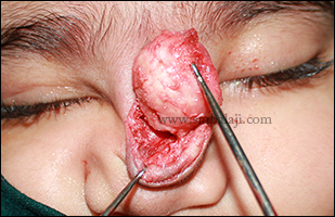
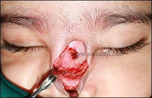
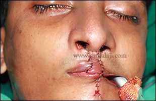
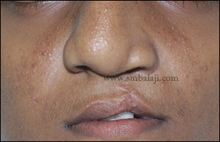
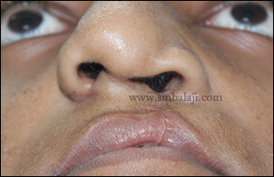
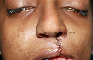
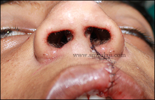
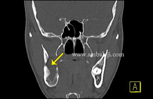
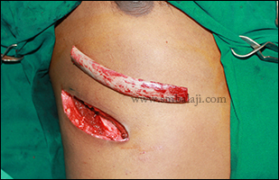
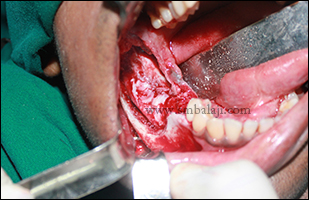
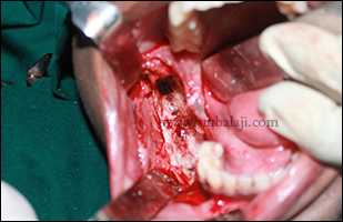
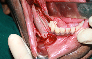
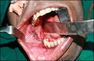
A 3D CT scan revealed temporomandibular joint ankylosis of the right side. Maxillofacial Surgeon Dr. S.M. Balaji successfully treated the jaw joint defect using gap arthroplasty with temporalis fascia interpositioning and contralateral coronoidectomy. Under GA, Al-Kayat-Bramley’s incision (Preauricular incision) was made on the right side; a superior limiting cut was made along the demarcation between the rim of the glenoid fossa and the ankylosed head. The ankylotic mass was removed. Intraoral coronoidectomy on the contralateral side i.e. the left side was done to facilitate good mouth opening. On the right side, the temporalis muscle was rotated and folded inwards into the joint cavity and taken between the medial surface of the ramus taking good care of the adjacent mandibular nerve, facial and maxillary arteries. Then the muscle end was sutured to the medial side of the mandible to provide a submandibular anchorage. A suction drain was placed and the wound was closed in layers. Following surgery mouth-opening improved considerably and the girl and her parents are very happy with the surgery outcome.
3D CT scan showing right side TMJ ankylosis- bony fusion of the condyle with base of skull
Severely restricted mouth opening and chin deviation
Temporalis muscle interpositioned between the glenoid fossa and condylat head and fixed
Greatly improved mouth opening seen immediately
Surgical site closed in layers and drain placed to prevent hematoma
Increased mouth opening following gap arthroplasty and temporalis interpositioning surgery
0 Comments

Successful correction of restricted eyelid movement due to orbital bone fracture

27/5/2015

0 Comments

 
A 30 year old man reported to our hospital with the complaints of inability to open the left eye completely due to fracture in the orbital bone due to road traffic accident 6 months back.

After thorough clinical and radiological examination, Maxillofacial Surgeon Dr. S. M. Balaji successfully corrected the orbital bone fracture without any visible surgical marks on the face. Through lid crease incision the fractured and displaced portion of superior orbital rim on the left side was reduced and fixed using 3hole stainless steel bone plate. Patient feels happy to have the entire correction done without any visible scar formation on the face.
Preoperative 3DCT scan view showing fractured and posterior displacement of lateral border of superior orbital rim
Intraoperative view showing displaced junction of lateral superior orbital rim
Fractured portion contoured and aligned to the orbital rim using 3 hole stainless steel plate
Immediately after suturing of the lid crease incision
0 Comments

Flat nose and disproportionate jaw correction without any scars on the face

21/5/2015

0 Comments

 
A 22 year old girl reported to our hospital wanting to correct her nose profile which was very broad and flat to her face and ill-defined dorsum gave her more aged appearance. On addition her prominent upper jaw on side view gave unsightly appearance. Due to her defective nose profile and disproportionate jaw alignment she was very self conscious about her appearance.

Maxillofacial Surgeon Dr. S. M. Balaji skillfully planned to correct her nose by following Augmentation Rhinoplasty technique. Costochondral graft was harvested from the ribs, shaped and used to reconstruct the nose to increase the height. Thus proper shape of the nose was achieved. Maxillary prominence was also corrected by following Lefort I osteotomy. Entire nose correction was made within the nose (closed rhinoplasty technique); there are no scars on the face. Results are immediate and the patient was very happy to see her enhanced profile of the nose without any scars. This dramatically improved her appearance and self confidence.
Preoperative facial view showing broad flat nose with ill defined nasal bridge
Profile view showing flat nose profile with maxillary protrusion
Costochondral rib graft harvested
Harvested rib graft used to augment the dorsum of the nose
Pristine nose without any visible scars on the face following a week after surgery
0 Comments

Combined lower jaw and chin correction for enhanced appearance

16/5/2015

0 Comments

 
A young man came to our hospital seeking corrective jaw surgery to enhance his appearance. His lower jaw was excessively protruding causing an incorrect teeth occlusion that caused difficulties in biting and chewing. Additionally his chin bone was deviated away from the midline giving his face an asymmetric, crooked appearance.
Maxillofacial surgeon Dr. S.M. Balaji planned to correct the jaw defect with a combined bilateral Obwegeser’s sagittal split osteotomy and genioplasty. The mandible was set back to correct the alignment and bite using bilateral Obwegeser’s sagittal split osteotomy. With genioplasty, the deviated chin bone was corrected. The procedure was done from inside the mouth so there were no scars. Immediately after this corrective jaw surgery, the man’s appearance was greatly improved and he is very happy with the enhanced aesthetics.
Preoperative profile view showing long lower jaw giving asymmetric profile
Preoperative 3DCT scan showing long lower jaw and reverse bite
Intraoperative view showing corrected occlusion by following bilateral sagittal split osteotomy with reduction genioplasty
Corrected facial profile following surgery without any scars on the face
0 Comments

Incomplete bilateral Cleft Lip- Successful Primary Correction

16/5/2015

0 Comments

 
A 4-month-old baby boy born with incomplete bilateral cleft lip and isolated cleft palate was brought to our hospital by his parents for specialized cleft lip & palate surgery.

Maxillofacial Surgeon Dr. S.M. Balaji performed the primary cleft lip correction surgery to reconstruct the lip enabling the baby to feed well while also dramatically enhancing the baby’s appearance.  Subsequent cleft palate surgery will be done to completely rehabilitate the child. 
Incomplete bilateral cleft lip in a 4 month old baby boy
Primary closure of incomplete cleft lip following Paul Black's technique
Immediately after suture removal following seven days of surgery
0 Comments

Cosmetic augmentation rhinoplasty without any scars

16/5/2015

0 Comments

 
A 24 year old girl reported to our hospital seeking cosmetic correction of her nose. She felt the dorsal surface of her nose was flat which gave her an aged look. She wanted to correct her nose profile cosmetically without any scar.

Maxillofacial Surgeon Dr. S. M. Balaji planned to correct her flattened nose by following closed augmentation rhinoplasty. Costochondral rib graft was harvested which was used to augment the dorsum of nose. To prevent scarring, the graft was inserted through intranasal approach. Bulky nose tip was corrected by removing caudal portion of nasal septal cartilage. Results are immediate, patient is very happy to have a sharp, pristine nose without any surgical marks, which suits her facial profile.
Preoperative facial view showing broad flat nose with ill defined nasal bridge and bulky tip
Costochondral rib graft harvested
Harvested rib graft used to augment the dorsum of the nose
Lower lateral nasal septal cartilage removed to reduce the bulky nose tip
Immediate postoperative view showing augmented dorsum of the nose giving pristine appearance without any visible surgical marks on the face
0 Comments

Augmentation rhinoplasty for saddle-shaped nose - Dr. S.M Balaji

14/5/2015

0 Comments

 
Surgical correction of nose deformities also known as the “Nose Job” requires expertise and experience.  The surgeons estimate the type of defect - either excess or deficit of the height, deviations, depressions, slants and abnormally angled noses. The patient’s expectation of his/her nose is understood after careful examination.

Shown is a case with deficiency of the bridge portion of nose. Note the midlevel of the nasal bone, where there is a deficiency creating a huge impact on the patient’s facial profile. This gives the nose a saddle-shape. Maxillofacial Surgeon Dr. S. M. Balaji planned a closed rhinoplasty technique wherein the surgical approach is through the nose to prevent unsightly scars.

Appropriate measurement of nose was made and the amount of augmentation needed was noted. Autologous graft (rib graft) was harvested, shaped and positioned to improve the aesthetics of the nose. The patient was very happy with the surgery outcome and his enhanced appearance.
Preoperative profile view of nose showing depressed dorsum with everted tip of the nose
Costochondral rib graft harvested to augment the dorsum of the nose
Intranasally the rib graft used to raise the height of the nose
Immediately after suturing augmented dorsum of nose without any visible surgical marks on the face
Postoperative profile view showing augmented dorsum of nose giving pristine appearance
0 Comments

Successful excision of Nasal Glial Heterotopia in a 9-month-old baby girl

12/5/2015

0 Comments

 
A 9-month-old baby girl from Orissa was brought to our hospital by her parents for removal of the large mass of tissue on the baby’s nose. The parents gave a history of the tissue mass appearing on the nose a few weeks after she was born. The nodule was initially small in size and later grew to the present size. The tissue mass on the nose was affecting the baby’s appearance and the parents were very worried about this abnormality.

Maxillofacial Surgeon Dr. S.M. Balaji successfully performed the excision biopsy. The entire mass of tissue was surgically excised from the base of the nodule to ensure complete removal. The surgical site was sutured. The baby’s facial appearance greatly improved after removal of the unsightly mass from the nose. A biopy of the mass tissue proved to be nasal glial heterotopia.
Facial view showing huge swelling on the dorsum of the nose in a 9 months old baby girl
Intraoperative view showing complete removal of the soft tissue swelling
Immediately after removal of the dermoid from the dorsum of the nose
Complete removal of the swelling in toto
Immediately after suturing
0 Comments

Successful cleft nose and lip correction with Abbe flap

9/5/2015

0 Comments

 
A 22 year old boy born with bilateral cleft lip and palate came to our hospital for expert cleft treatment. He was operated elsewhere for primary cleft lip and palate. He had a thin upper lip and broad flattened nose for which he sought treatment.

Maxillofacial Surgeon Dr. S.M. Balaji performed the specialized Abbe flap technique to correct the upper lip. A flap of tissue was taken from the lower lip and used to reconstruct the upper lip with the base of the flap still attached to the lower lip. Once blood supply is established, the flap will be divided giving the upper lip a more natural, improved form.

Simultaneously, the broad nose was reshaped to make them more proportionate and balanced. Following surgery, the nose appeared sharp & defined with more proportionate profile.
Preoperative view showing broad flat nose with defective upper lip
Profile view showing ill defined columella of nose with depressed nasal tip and defective upper lip
Intraoperative view showing raised columella of nose and correction of upper cleft lip defect
Immediately after suturing showing corrected nose and lip defect using Abbe flap technique
Postoperative profile view showing raised dorsum of nose with nasal tip giving pristine appearance
0 Comments

Cleft rhinoplasty without scars - Dr. S.M Balaji

5/5/2015

0 Comments

 
A 15-year-old boy reported to our hospital seeking specialized treatment to correct his cleft nose defect. He was previously operated for cleft lip and palate elsewhere. Due to the cleft defect his nose was collapsed on the left side and it affected his facial appearance.

Maxillofacial Surgeon Dr. S.M. Balaji expertly performed the rhinoplasty or nose correction. A costochondral graft (rib graft) was harvested and used to reconstruct the collapsed nasal framework. The surgery was approached from inside the nose so there was no scar. Secondary lip repair was also done to correct the vermillion defect. Immediately after surgery, the nose appeared sharp and definite giving a more pleasing appearance. The patient was very happy to have his nose defect corrected without any visible surgical marks on the face.
Preoperative facial view showing broad nose with deviated nose tip towards right side due to cleft defect
Columellar view showing depressed nasal tip with ill defined columella and asymmetric nostrils
Costochondral rib graft harvested to reconstruct the shape of the nose
Immediately after suturing showing corrected dorsum of the nose with cleft lip revision
Columellar view showing corrected shape of the nostrils using rib graft intranasally without any visible surgical marks on the face
0 Comments

Successful reconstruction of infected portion of mandible due to sequestrum of failed bovine bone grafting done elsewhere

2/5/2015

0 Comments

 
A 30 year old man came to our hospital with the complaints of swelling in right side of face with pain for past 3 months. He gave the history that he had a huge osteolytic lesion involving right side of the mandible which was removed and reconstructed with bovine bone grafting elsewhere. 

Due to graft rejection there was a huge infected cyst with sequestrum (Dead bone) in the involved region of mandible. Intraorally there was an incomplete closure of buccal mucosa where the sequestrum was exposed. After thorough clinical and radiological examination, Maxillofacial Surgeon Dr. S. M. Balaji successfully removed the dead bone with the infected cyst intraorally and reconstructed the same with costochondral rib graft. Patient feels happy to have both the removal and reconstruction of the infected portion of mandible without any visible surgical marks on the face.
Preoperative 3DCT scan view showing complete destruction of ramus of mandible on right side with infected cyst
Harvested costochondral rib graft used to reconstruct the defective portion of mandible
Infected portion of mandible surgically exposed showing sequestrum of the failed bovine bone graft done elsewhere
After complete removal of the cyst along with sequestrum of the bovine bone graft
Harvested rib graft used to reconstruct the defective portion of mandible
Immediately after suturing
0 Comments

    Categories

    All
    Abbe Flap Technique
    Advanced Transport Distraction Osteogenesis
    Advancement Genioplasty
    Alveolar Cleft Repair
    Alveolar Distraction
    Ameloblastoma
    Augmentation Genioplasty
    Augmentation Rhinoplasty
    Bat Ear Correction
    Bilateral Cleft Lip Palate
    Blepharoplasty
    Bone Defect Reconstruction
    Bone Grafting
    Bone Morphogenetic Protein(BMP)
    Broad Flattened Nose Correction
    Bulky Nose Correction
    Chin Correction
    Chin Correction Surgery
    Cleft Lip & Palate Surgery
    Cleft Nose
    Cleft Palate Repair
    Cleft Rhinoplasty
    Complete Facial Rehabilitation
    Complex Facial Fractures
    Condylar Fracture Surgery
    Correction Of Hypoplastic Ramus
    Corrective Jaw Surgery
    Cosmetic Cleft Nose Correction
    Cosmetic Nose Correction
    Cosmetic Rhinoplasty
    Cosmetic Surgery
    Craniofacial Deformities
    Craniofacial Reconstruction
    Craniofacial Surgery
    Cyst Removal
    Dental Implant
    Dentigerous Cyst
    Deviated Nose Correction
    Diplopia
    Distraction Osteogenesis
    Ear Correction
    Ear Defect
    Ear Reconstructive Surgery
    Epiphora
    Extruding Infected MEDPOR
    Eyebrow Correction
    Face Enhancement
    Face Reconstruction
    Facial Asymmetry Correction
    Facial Bone Fracture
    Facial Burn Injury
    Facial Palsy Surgery
    Fibrous Dysplasia
    Flat Nose Correction
    Forehead Bone Fracture
    Forehead Deformity
    Forehead Flap
    Frontal Bossing Correction
    Gummy Smile
    Hemifacial Microsomia
    Hypertelorism Surgery
    Implant
    Incomplete Cleft Lip
    Isolated Cleft Palate Repair
    Jaw Correction Surgery
    Jaw Cyst
    Jaw Deformity
    Jaw Fracture Surgery
    Jaw Joint Ankylosis
    Jaw Reconstructive Surgery
    Jaw Surgery
    Lip Correction
    Lip Defect Correction
    Lip Revision
    Locked Jaw
    Lower Jaw Fracture
    Low Lying Upper Eyelid Correction
    Macrostomia Correction
    Mandible Correction
    Mandible Reconstruction
    Maxillofacial Surgery India
    Maxillomandibular Distraction
    Maxilofacial Surgery
    Micrognathic Mandible Correction
    Microtia
    Nasal Glial Heterotopia
    Naso-orbito-ethmoidal Fracture
    Nose And Lip Correction
    Nose Asymmetry Correction
    Nose Correction
    Nose Job
    Nose Reconstructive Surgery
    Oral Submucous Fibrosis
    Oral Tumor Removal
    Orbital Correction
    Orehead Defect
    Orthognathic Surgery
    Parrot-beak Nose
    Parry Romberg Syndrome
    Pharyngoplasty
    Plastic Surgery
    Primary Cleft Lip Repair
    Profile EnhancemeP
    Reduction Glossectomy
    Reduction Rhinoplasty
    Removal Of Capillary Hemangioma
    RhBMP 2
    RhBMP-2
    Rhinoplasty
    Road Accident Trauma
    Road Accident - Trauma
    Scar Revision
    Secondary Cleft Palate
    Sinus Lift
    Smile Makeover
    Speech Correction
    Square-face Correction
    Surgery Without Scar
    Tessier Nose Defect
    Tissue Expansion
    TMJ Ankylosis
    Tongue Correction
    Tongue Surgery
    Trauma
    Trauma Treatment
    Triangular Shaped Forehead
    Tumor Resection
    Unilateral Cleft Lip
    Unilateral Cleft Lip And Palate
    Unilateral Cleft Lip Repair
    Velopharyngeal Incompetence

    Archives

    April 2021
    March 2021
    January 2021
    December 2020
    April 2020
    March 2019
    February 2019
    January 2018
    November 2017
    October 2017
    September 2017
    June 2017
    May 2017
    April 2017
    March 2017
    January 2017
    December 2016
    October 2016
    September 2016
    August 2016
    July 2016
    June 2016
    May 2016
    April 2016
    March 2016
    February 2016
    January 2016
    December 2015
    October 2015
    September 2015
    August 2015
    July 2015
    June 2015
    May 2015
    April 2015
    March 2015
    February 2015
    January 2015
    December 2014
    November 2014
    October 2014
    September 2014
    August 2014
    July 2014
    June 2014
    May 2014
    April 2014
    March 2014
    February 2014
    January 2014
    December 2013
    November 2013

    RSS Feed

Copyright © 2020 Balaji Dental & Craniofacial Hospital | All rights reserved | Powered by BALAJI DENTAL HOSPITAL