
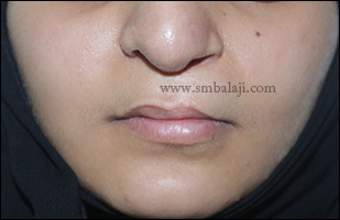
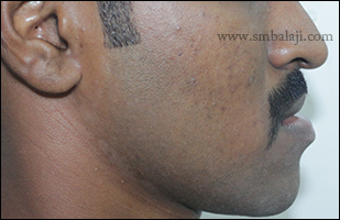
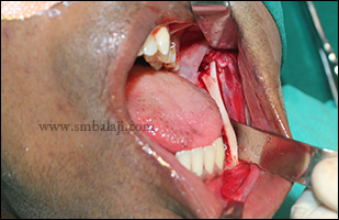
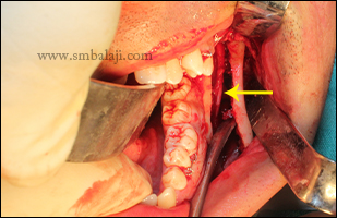
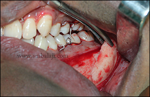
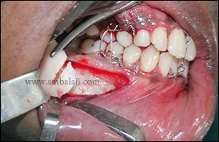
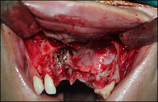
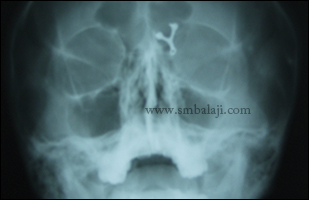
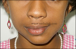
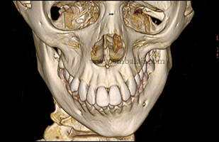
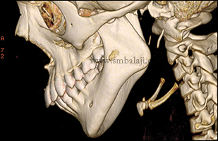
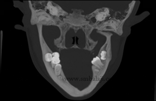
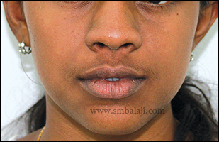
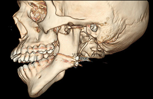
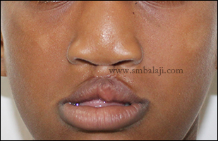
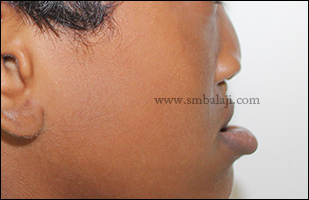
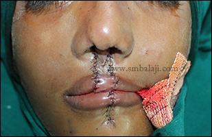
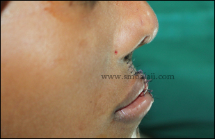
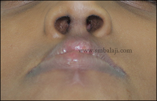
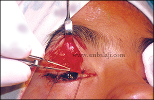
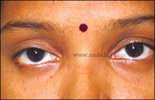
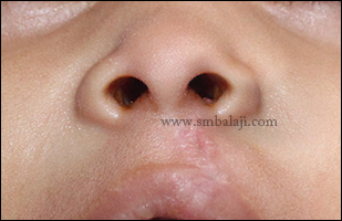
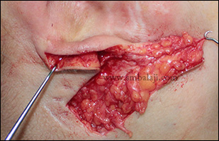
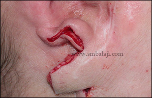
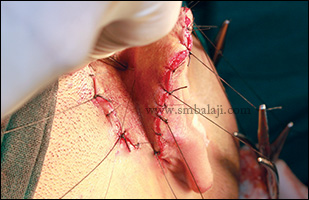
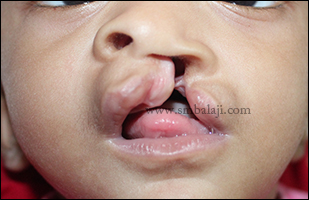
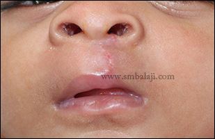
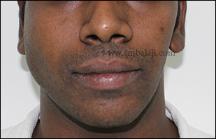
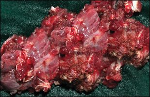
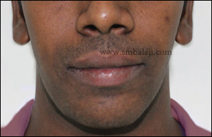
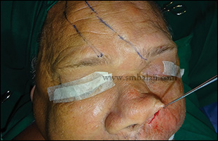
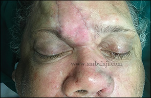
A 24 year old girl from Dubai, a known case of unilateral cleft lip and palate reported to our hospital seeking for lip correction. Primary cleft lip repair was done elsewhere resulting in thinning of upper lip. Maxillofacial Surgeon Dr. S. M. Balaji successfully corrected this defect using bullhorn lip lift technique.
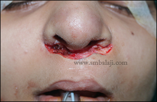
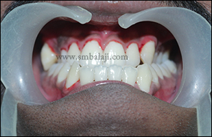
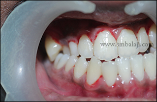
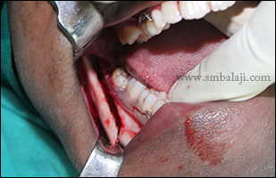
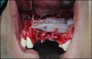
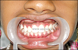
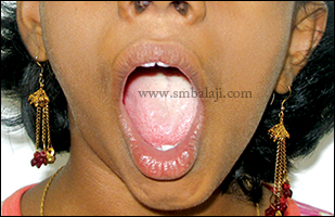
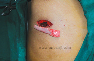
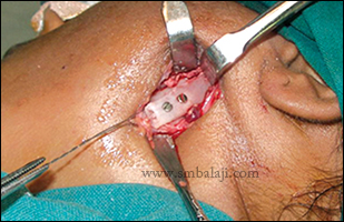
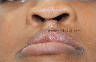
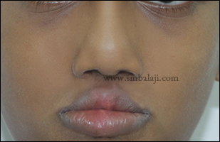
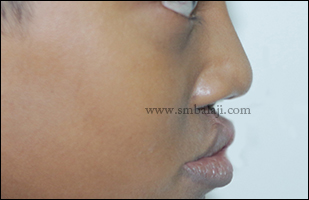
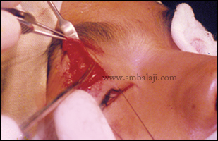
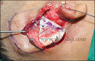
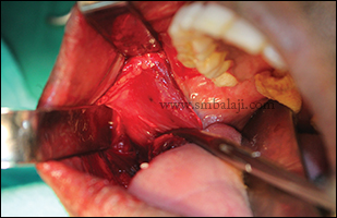
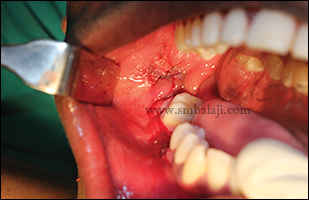
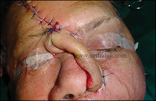
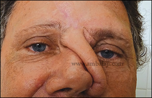
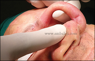
There are different types of lip lift surgery. The most commonly used lip lifting technique is known as the ‘bullhorn lip lift’. Pre-operative markings on the skin ensure the lip lift is approximately symmetric. Advancement of the inferior edge of skin directly beneath the nasal base lifts the lip, producing more visible vermilion and about 3 mm of teeth show at rest. The position of the final incision is such that it is located within the shadow of the nose. Meticulous technique produces an almost invisible scar. The amount and width of skin excised is individualized depending on the desired aesthetic goals. Lip lifts using this technique provide an immediate, dramatic, and permanent result.
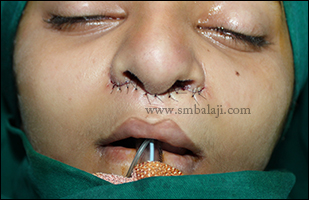
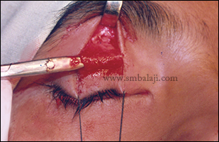
There are different types of lip lift surgery. The most commonly used lip lifting technique is known as the ‘bullhorn lip lift’. Pre-operative markings on the skin ensure the lip lift is approximately symmetric. Advancement of the inferior edge of skin directly beneath the nasal base lifts the lip, producing more visible vermilion and about 3 mm of teeth show at rest. The position of the final incision is such that it is located within the shadow of the nose. Meticulous technique produces an almost invisible scar. The amount and width of skin excised is individualized depending on the desired aesthetic goals. Lip lifts using this technique provide an immediate, dramatic, and permanent result.


 RSS Feed