Best Dental Clinic in Chennai : Best Dental Implant Center in India
FOLLOW US
  • HOME
  • Surgery of The week
  • Clinical Updates
  • Archives
  • Latest News
  • VIDEOS
  • CONTACT US

Facial asymmetry correction using Distraction osteogenesis

13/5/2016

0 Comments

 
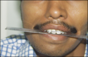
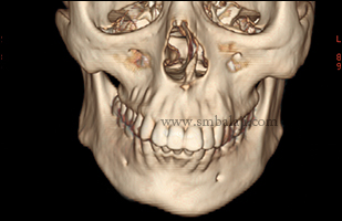
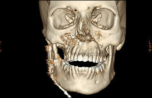
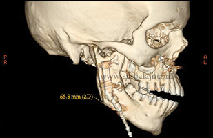
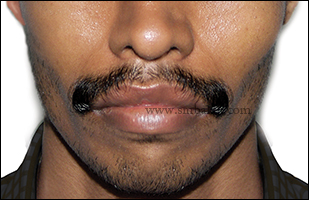
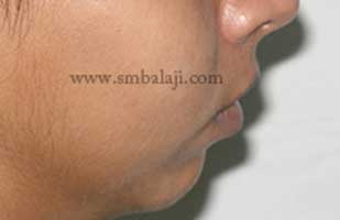
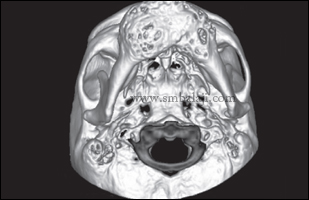
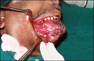
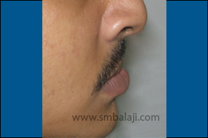
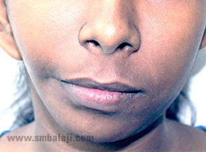
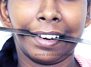
Hemifacial microsomia is a condition in which there is an under-development of one side of the face resulting in facial asymmetry. The affected side of the face appears disproportionately smaller than the other. Characteristically, the lower jaw (mandible), facial soft tissues & musculature, cheek and ear on one side of the face is poorly developed. Sometimes, structural defects in the eye may also be seen. A 28-year-old boy afflicted with Hemifacial Microsomia reported to our Hospital. His lower jaw and ear was under-developed on the right side. He had a slanting bite and an obvious asymmetry of the face. A radiographic examination showed a grossly under-developed lower jaw (mandible) on the right side. There was found to be a bone deformity of about 20 mm.
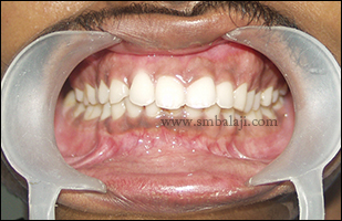
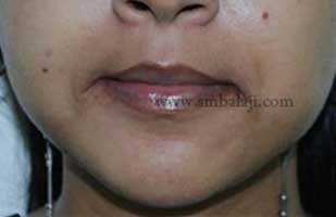
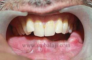
The goal of the treatment is to elongate the deficient jaw bone to restore facial symmetry and correct the slanting bite (occlusion). To achieve this, an advanced and effective treatment technique is distraction osteogenesis. This is a new technique for regenerating new bone by slow, progressive stretching of the bone, without requiring a bone graft.
Eminent Craniofacial Surgeon Dr. S.M. Balaji is a pioneer in introducing this revolutionary technique and has successfully rehabilitated the maximum number of patients afflicted with facial disfigurements in the country.
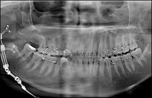
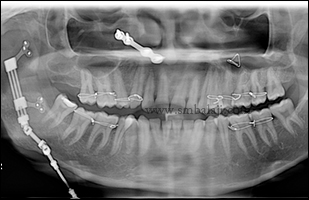
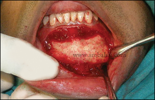
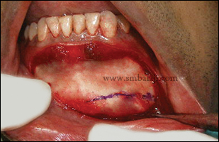
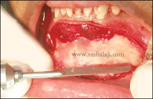
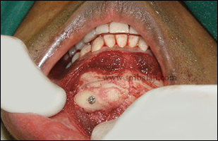
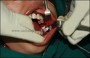
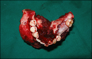
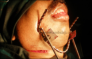
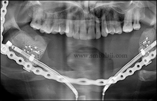
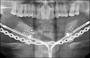
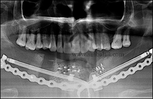
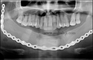
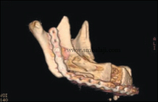
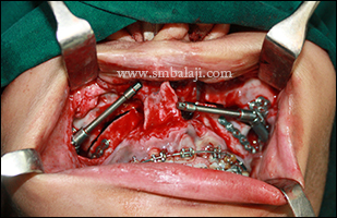
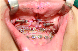
In distraction osteogenesis, the jaw bone on the deficient side is cut. A sophisticated device called distractor is placed such that the two arms of the device are fixed to the two segments of jaw bone. After a few days, a screw attached to the distractor is turned gradually, ideally at a rate of 1 mm per day. When this is done, the two cut segments move apart and new bone is formed in the resultant gap. After the required length is achieved and new bone is stabilized, the distractor device is removed subsequently correcting the asymmetry of the face.
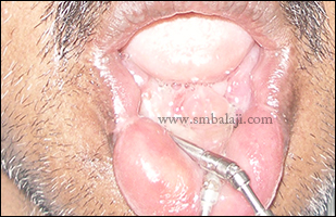
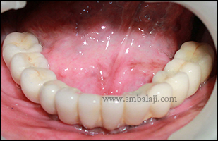
This is a powerful tissue engineering technique for generating bone for the desired volume. The overlying soft tissues that are deficient also are expanded, therefore along with the underlying bone deformity, the overlying skin and soft tissue defect is also corrected.
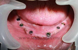
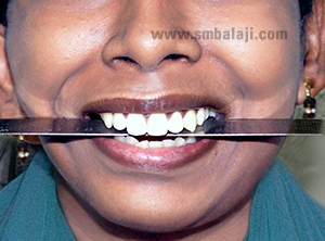
This is the only procedure to increase the size of the jaw bones after the cessation of actual bone growth. For this patient, Dr. Balaji adopted a stage-by-stage rehabilitation. To correct the jaw deformity, he applied the innovative simultaneous maxillary and mandibular distraction procedure wherein new bone is formed in both the upper and lower jaws simultaneously to restore facial symmetry.
Preoperative facial view showing asymmetrical face ( short face on left side)
Slanting bite (occlusal cant) due to retarded development of facial bones on left side
Preoperative 3DCT showing retarded development of maxilla and mandible on left side
After distraction showing symmetrical appearance and elongated genium highlighted in 3DCT scan image
Immediately after fixing the distractor device in position
After completion of distraction and formation of callus at the end of consolidation before distractor removal
3DCT scan showing good new bone formation
Enhanced bone formation following distraction osteogenesis
During distractor removal the genial excess was alos corrected
Excess bone was highlighted
Excess bone was removed
Excess bone removed was used to augment the symphysis on other side
Postoperative facial view showing symmetrical face without any scars on the face
Postoperative occlusal view showing corrected bite
0 Comments

Micrognathic mandible correction using simultaneous internal maxillomandibular distraction osteogenesis

17/2/2016

0 Comments

 
22 year old female, a known case of bilateral TMJ ankylosis reported to our hospital for the surgical correction of lower half of the face. Micrognathic mandible present due to retarded development. She gave the history of TMJ ankylosis release surgery done elsewhere during childhood.
After thorough clinical and radiological examination Maxillofacial Surgeon Dr. S. M. Balaji planned to correct the facial asymmetry using internal distraction osteogenesis with maxillomandibular distractor. Bilaterally Paragingival incision was placed over the angle region along the anterior border of ramus. Full thickness mucoperiostal flap was reflected buccally and bone exposed. The direction of the osteotomy cut and positioning of the distractor is the most important as it determines the vector of distraction and this determines the direction of the bone growth. The distractor device was positioned and fixed using screws. Osteotomy is then completed along the medial cortex. Distractor device was checked and wound closure done. Le Fort I osteotomy was completed in maxilla.
After a latency period of 5 days, distraction at the rate of 1 mm per day was accomplished and facial asymmetry was successfully corrected. Thus maxillomandibular distraction technique helped dramatically to enhance the facial appearance without any visible scars and taking autobone graft anywhere from the body. Patient feels very happy to have enhanced facial appearance without any scar on the face.
Retruded mandible due to bilateral temporomandibular joint ankylosis
Retruded mandible with illdefined chin prominence
Intraoperative view showing intraoral fixation of maxillomandibular distractors
Postoperative view after distraction shows forwardly moved mandible without any visible scars on the face
Postoperative profile view showing advanced lower jaw with enhanced chin prominence
0 Comments

Advanced transport Distraction osteogenesis technique for reconstruction of lower jaw after tumor removal

11/2/2016

0 Comments

 
A 55 year old patient reported to our hospital with a large, bony swelling in the anterior portion of the lower jaw (mandible). He had said that the swelling had gradually increased in size over the past 1 year and there was spontaneous loss of few lower teeth over the last 6 months. He also reported of altered sensation in the lower lip for past 3 weeks which was his main concern.
​
 A 3D CT scan was taken to gauge the extent of the lesion. A biopsy was done which showed the lesion to be a multiple cystic ameloblastoma (tumor) of the lower jaw. Maxillofacial Surgeon Dr. S. M. Balaji removed the entire bony tumor along with the involved bone and reconstructed the defective portion of mandible using the native bone itself without any bone graft by following advanced transport guided distraction osteogenesis technique.

The entire affected portion of the lower jaw was ressected with 1.5 cm marginal clearance followed by reconstruction using titanium plate guided bilateral tripod multi docking complex univector Herford plate guided mandibular transport distractor.

After 5 days of latency period, the distractors were activated at a rate of 0.5mm twice per day (on both sides) until the transport discs had reached the canine region on both sides (amount of distraction on the right side was 46mm and on the left 43.2mm). Following consolidation period of 3 months and radiological evidence of corticalization, the distractors were removed. And complete dental implant rehabilitation done for lower arch in the newly formed healthy bone. Cosmetically well blended fixed ceramic prosthesis given.
X-ray showing tumor in the lower jaw
3DCT scan view showing huge tumor involving more than half of the lower jaw
Intraoperative view showing removal of huge tumor mass intraorally
Complete removal of the tumor mass along with the involved bone in toto
Planning for the fixation of the transport distractor device to the mandible
After tumor removal, fixation of reconstruction plate and segments distracted 1 mm per day
During transport distraction
During distraction
After completion of distraction
After completion of the distraction - facial view
After removal of the distractors and fixing the fragments to the reconstruction plate to achieve the 'U' form of the lower jaw
Healthy new bone formation seen in postoperative 3DCT scan
Postoperative intraoral view showing enhanced healing and augmented new alveolar bone formation
Implants placed in the reconstructed lower jaw
Following dental implant rehabilitation cosmetically well blended fixed ceramic prosthesis placed
0 Comments

Correction of defective upper jawbone with internal maxillary distraction osteogenesis

2/1/2015

0 Comments

 
This is a case of unilateral cleft lip and palate operated elsewhere. His profile view was asymmetric due to defective upper jaw bone which also developed difficulty in speech and chew foods. Maxillofacial Surgeon Dr. S. M. Balaji successfully corrected the defective upper jawbone by using intraoral bilateral maxillary distraction osteogenesis technique following orthodontic treatment to have perfect occlusion postoperatively. Results are immediate and patient is very happy of his improved appearance and to have entire jaw correction done without any scars.
Preoperative profile view showing defective upper jaw due to cleft defect
Intraoperative view showing lefort I osteotomy with bilateral maxillary internal distractor fixed
Intraoperative view showing internal maxillary distractor fixation following orthodontic treatment
Postoperative view following internal maxillary distraction osteogenesis giving enhanced appearance
0 Comments

Facial asymmetry correction with advanced distraction osteogenesis technique

16/6/2014

0 Comments

 
A 20 year old girl reported to our hospital with a complaint of asymmetrical face giving her an unpleasant appearance and she was not able to chew foods due to slanting bite. Her parents were also worried & wanted the correction done without any scars on the face. The girl was too anxious due to her asymmetrical face which affected her self-esteem considerably.

After thorough clinical and radiological examination, it was diagnosed that she had hemifacial microsomia in which the ramul height of the mandible on right side was considerably less which led to asymmetry. Maxillofacial Surgeon    Dr. S. M. Balaji skillfully planned to correct her face as well as her bite with the innovative “Distraction Osteogenesis” technique. Intraorally Le Fort I osteotomy was done, lower jaw bone on the right side was cut and distractor device was fixed to the mandible at the ramus region. The screw attached to the distractor was turned gradually everyday that pushed the cut bone segments apart. New bone was formed in the resultant gap and the jaw bone was ultimately lengthened. After the face became symmetrical, the distractor was removed.

Thus entire surgery was done inside the mouth with no scars on the face.  The girl and her parents were very happy with the results.
Preoperative view showing facial asymmetry due to reduced height of the lower jaw on right side
Preoperative view showing slanting bite towards right side due to hemifacial defect
Distractor fixed in the right side of the mandible to increase the height of the jaw
Postoperative view showing enhanced appearance of the face with symmetrical appearance
Postoperative view showing corected bite with symmetrically aligned upper and lower jaw without any scars on the face
0 Comments

    Categories

    All
    Abbe Flap Technique
    Advanced Transport Distraction Osteogenesis
    Advancement Genioplasty
    Alveolar Cleft Repair
    Alveolar Distraction
    Ameloblastoma
    Augmentation Genioplasty
    Augmentation Rhinoplasty
    Bat Ear Correction
    Bilateral Cleft Lip Palate
    Blepharoplasty
    Bone Defect Reconstruction
    Bone Grafting
    Bone Morphogenetic Protein(BMP)
    Broad Flattened Nose Correction
    Bulky Nose Correction
    Chin Correction
    Chin Correction Surgery
    Cleft Lip & Palate Surgery
    Cleft Nose
    Cleft Palate Repair
    Cleft Rhinoplasty
    Complete Facial Rehabilitation
    Complex Facial Fractures
    Condylar Fracture Surgery
    Correction Of Hypoplastic Ramus
    Corrective Jaw Surgery
    Cosmetic Cleft Nose Correction
    Cosmetic Nose Correction
    Cosmetic Rhinoplasty
    Cosmetic Surgery
    Craniofacial Deformities
    Craniofacial Reconstruction
    Craniofacial Surgery
    Cyst Removal
    Dental Implant
    Dentigerous Cyst
    Deviated Nose Correction
    Diplopia
    Distraction Osteogenesis
    Ear Correction
    Ear Defect
    Ear Reconstructive Surgery
    Epiphora
    Extruding Infected MEDPOR
    Eyebrow Correction
    Face Enhancement
    Face Reconstruction
    Facial Asymmetry Correction
    Facial Bone Fracture
    Facial Burn Injury
    Facial Palsy Surgery
    Fibrous Dysplasia
    Flat Nose Correction
    Forehead Bone Fracture
    Forehead Deformity
    Forehead Flap
    Frontal Bossing Correction
    Gummy Smile
    Hemifacial Microsomia
    Hypertelorism Surgery
    Implant
    Incomplete Cleft Lip
    Isolated Cleft Palate Repair
    Jaw Correction Surgery
    Jaw Cyst
    Jaw Deformity
    Jaw Fracture Surgery
    Jaw Joint Ankylosis
    Jaw Reconstructive Surgery
    Jaw Surgery
    Lip Correction
    Lip Defect Correction
    Lip Revision
    Locked Jaw
    Lower Jaw Fracture
    Low Lying Upper Eyelid Correction
    Macrostomia Correction
    Mandible Correction
    Mandible Reconstruction
    Maxillofacial Surgery India
    Maxillomandibular Distraction
    Maxilofacial Surgery
    Micrognathic Mandible Correction
    Microtia
    Nasal Glial Heterotopia
    Naso-orbito-ethmoidal Fracture
    Nose And Lip Correction
    Nose Asymmetry Correction
    Nose Correction
    Nose Job
    Nose Reconstructive Surgery
    Oral Submucous Fibrosis
    Oral Tumor Removal
    Orbital Correction
    Orehead Defect
    Orthognathic Surgery
    Parrot-beak Nose
    Parry Romberg Syndrome
    Pharyngoplasty
    Plastic Surgery
    Primary Cleft Lip Repair
    Profile EnhancemeP
    Reduction Glossectomy
    Reduction Rhinoplasty
    Removal Of Capillary Hemangioma
    RhBMP 2
    RhBMP-2
    Rhinoplasty
    Road Accident Trauma
    Road Accident - Trauma
    Scar Revision
    Secondary Cleft Palate
    Sinus Lift
    Smile Makeover
    Speech Correction
    Square-face Correction
    Surgery Without Scar
    Tessier Nose Defect
    Tissue Expansion
    TMJ Ankylosis
    Tongue Correction
    Tongue Surgery
    Trauma
    Trauma Treatment
    Triangular Shaped Forehead
    Tumor Resection
    Unilateral Cleft Lip
    Unilateral Cleft Lip And Palate
    Unilateral Cleft Lip Repair
    Velopharyngeal Incompetence

    Archives

    April 2021
    March 2021
    January 2021
    December 2020
    April 2020
    March 2019
    February 2019
    January 2018
    November 2017
    October 2017
    September 2017
    June 2017
    May 2017
    April 2017
    March 2017
    January 2017
    December 2016
    October 2016
    September 2016
    August 2016
    July 2016
    June 2016
    May 2016
    April 2016
    March 2016
    February 2016
    January 2016
    December 2015
    October 2015
    September 2015
    August 2015
    July 2015
    June 2015
    May 2015
    April 2015
    March 2015
    February 2015
    January 2015
    December 2014
    November 2014
    October 2014
    September 2014
    August 2014
    July 2014
    June 2014
    May 2014
    April 2014
    March 2014
    February 2014
    January 2014
    December 2013
    November 2013

    RSS Feed

Copyright © 2020 Balaji Dental & Craniofacial Hospital | All rights reserved | Powered by BALAJI DENTAL HOSPITAL