
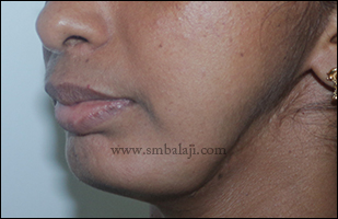
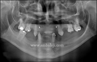
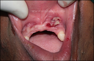
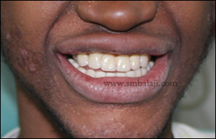
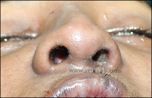
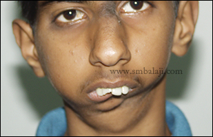
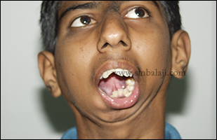
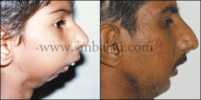
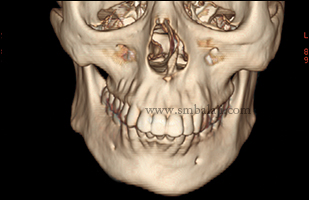
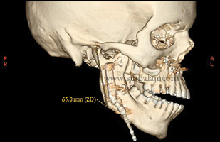
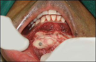
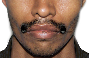
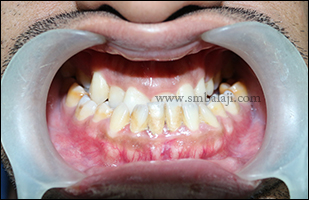
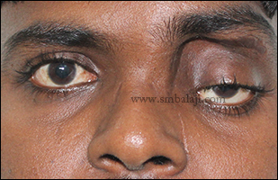
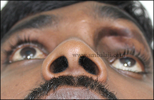
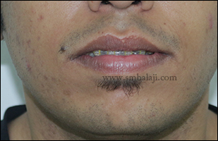
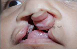
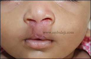
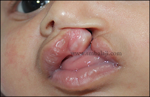
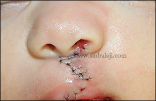
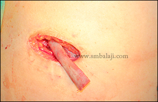
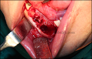
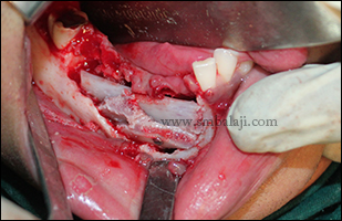
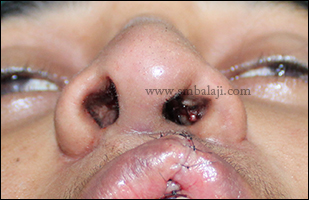
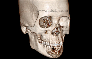
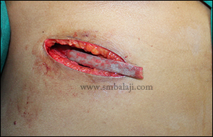
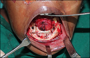
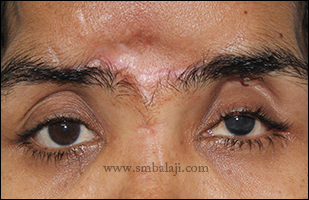
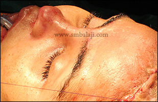
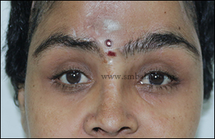
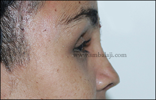
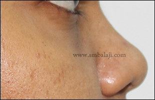
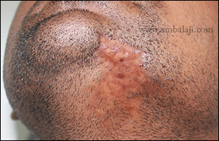
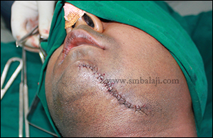
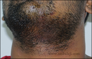
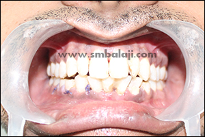
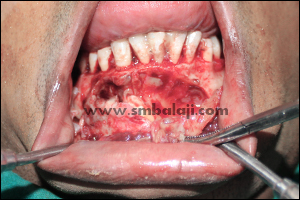
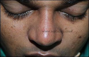
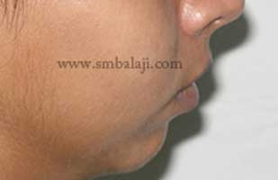
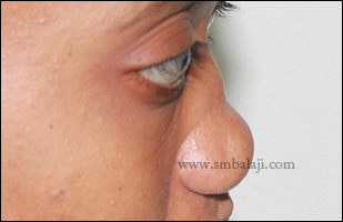
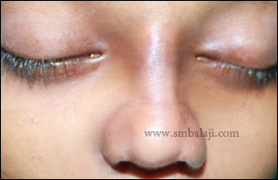
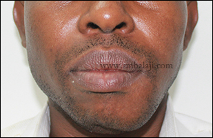
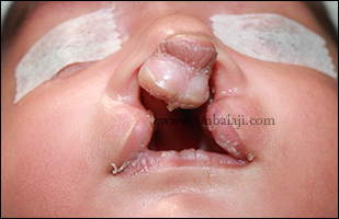
A 32 year old woman came to our hospital for expert treatment for her lower jawbone defect. She gave the history of huge odontogenic keratocyst lesion involving left mandibular angle and ramus along with the entire condylar component was removed extraorally and reconstructed using costochondral rib graft elsewhere. But the graft got infected and removed and reconstruction plate was fixed. Later plate was broken and was removed. Her complaint was jaw deviation towards left side and facial asymmetry. Also she wants to correct her facial appearance without any scars on the face.
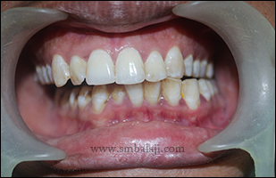
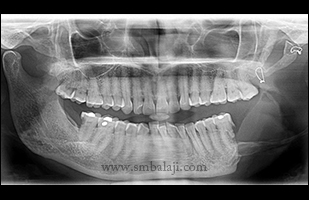
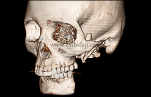
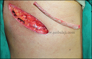
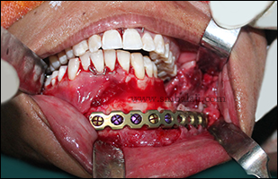
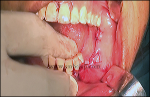
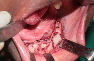
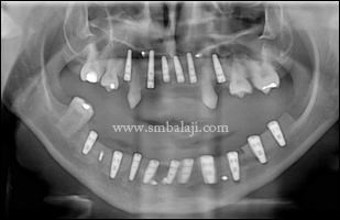
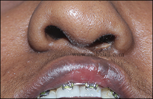
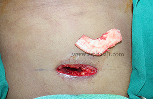
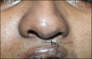
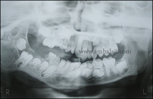
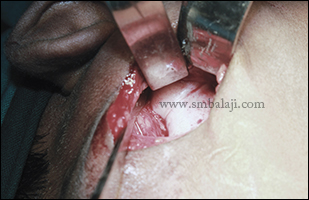
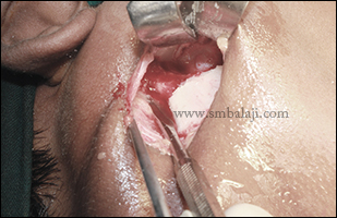
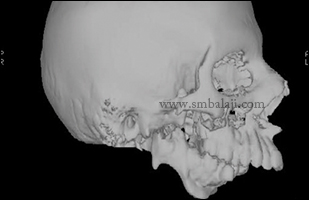
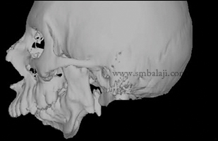
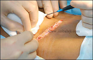
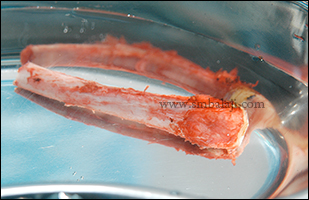
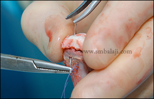
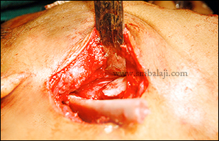
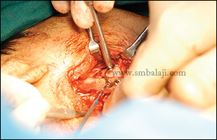
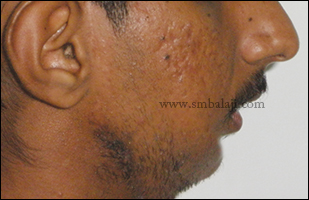
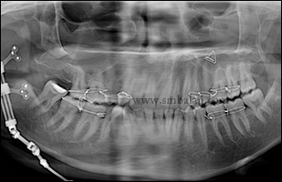
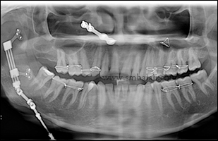
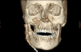
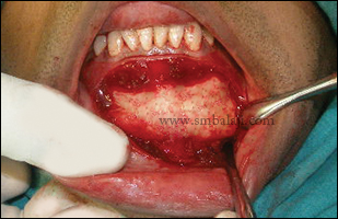
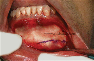
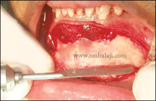
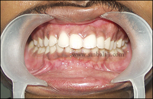
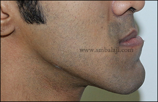
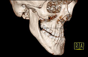
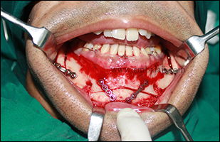
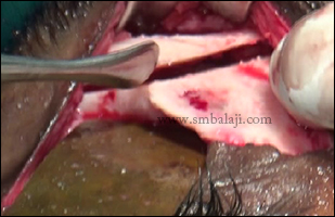
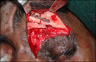
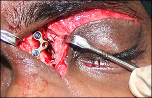
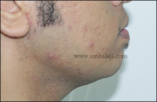
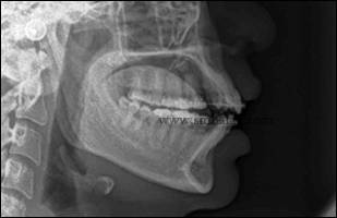
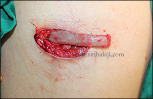
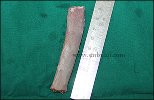
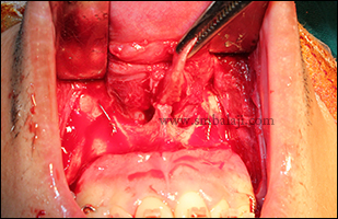
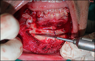
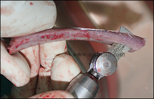
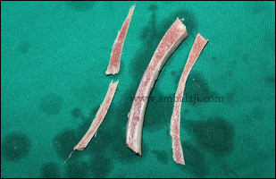
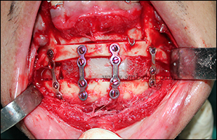
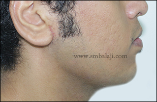
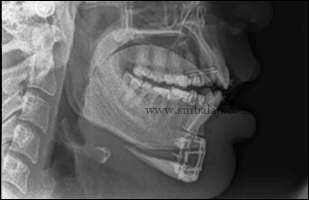
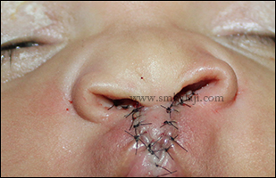
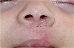
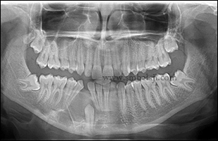
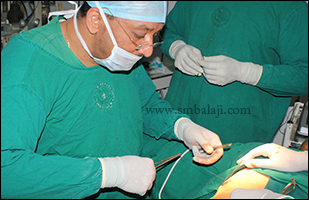
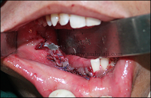
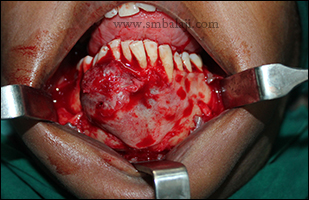
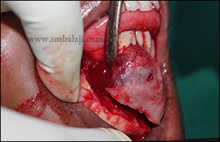
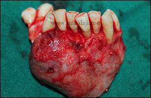
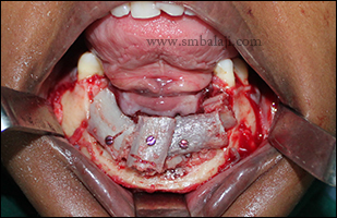
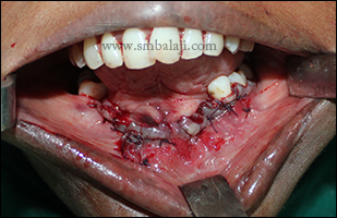
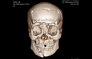
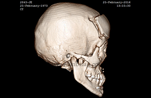
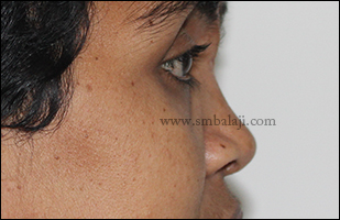
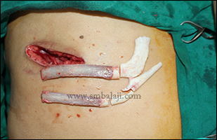
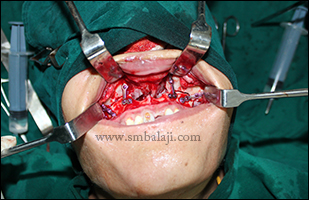
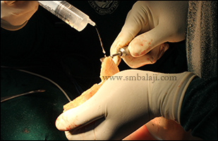
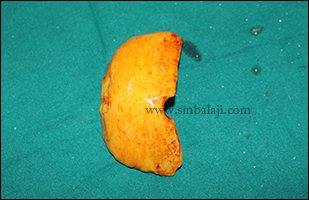
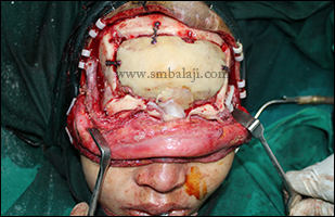
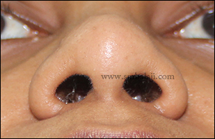
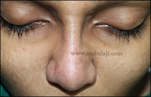
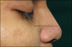
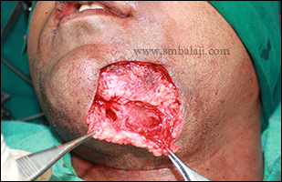
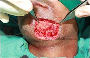
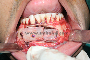
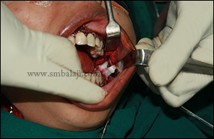
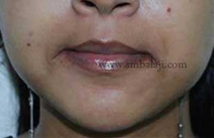
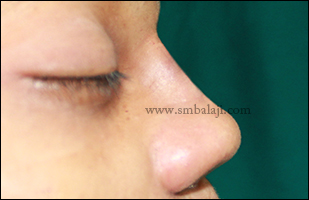
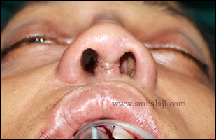
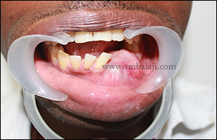
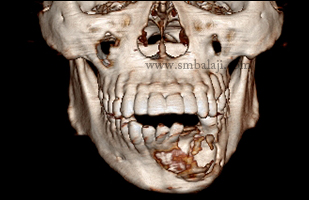
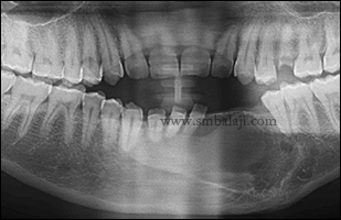
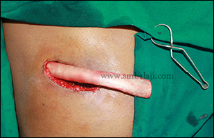
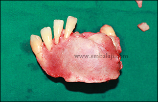
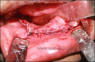
Maxillofacial Surgeon Dr. S.M. Balaji performed the clinical and radiological investigations. OPG and 3D CT scan showed complete absence of left side angle and ramus of mandible with remnants of bone graft. Dr. Balaji successfully reconstructed the huge mandibular defect maintaining the contour using titanium “L” plate and costochondral rib graft and thus missing condylar component was also reconstructed, the surgical site was closed. Patient feels very happy to have entire surgery done inside the mouth and to have complete facial asymmetry correction without any scars on the face.
Maxillofacial Surgeon Dr. S.M. Balaji performed the clinical and radiological investigations. OPG and 3D CT scan showed complete absence of left side angle and ramus of mandible with remnants of bone graft. Dr. Balaji successfully reconstructed the huge mandibular defect maintaining the contour using titanium “L” plate and costochondral rib graft and thus missing condylar component was also reconstructed, the surgical site was closed. Patient feels very happy to have entire surgery done inside the mouth and to have complete facial asymmetry correction without any scars on the face.


 RSS Feed