
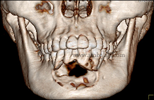
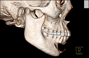
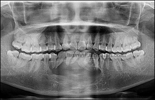
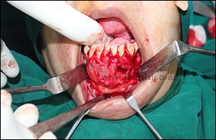
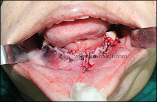
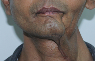
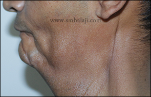
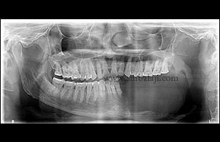
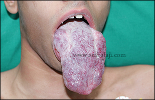
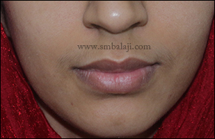
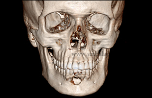
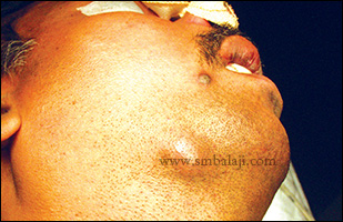
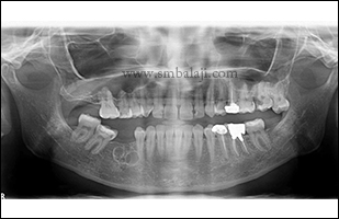
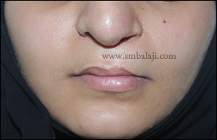
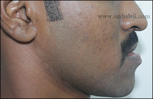
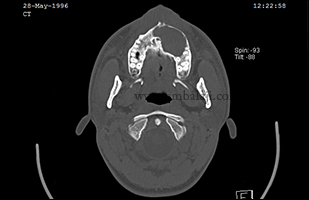
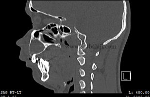
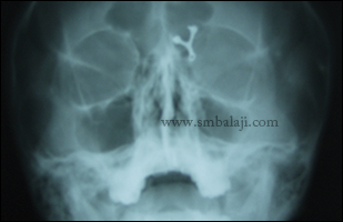
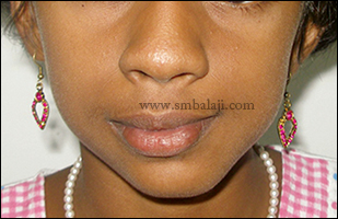
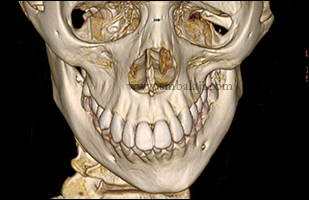
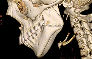
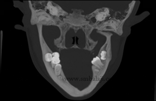
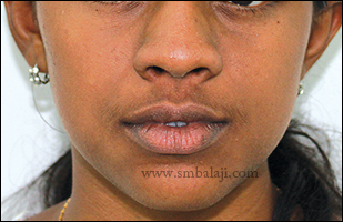
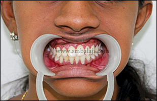
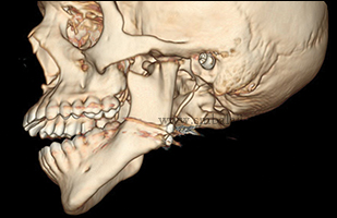
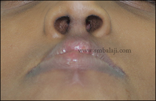
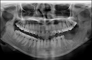
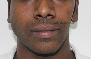
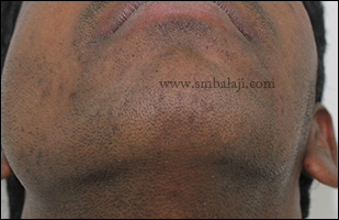
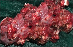
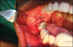
A 32 year old woman reported to our hospital with the complaint of swelling with pain in the front region of lower jaw. She added that the pain was only for last 3 months and the swelling was gradually increasing to the present size for a period of 6 months.
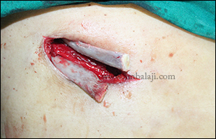
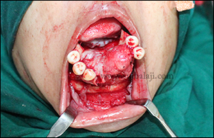
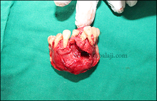
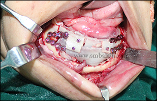
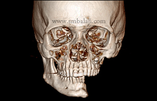
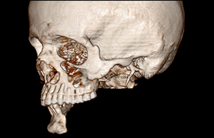
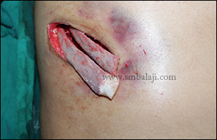
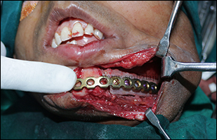
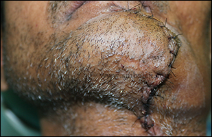
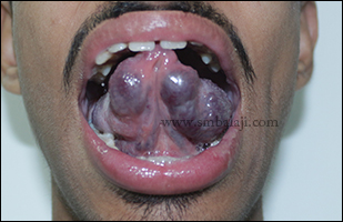
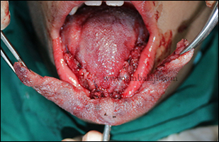
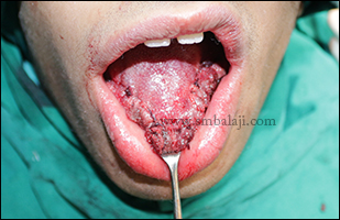
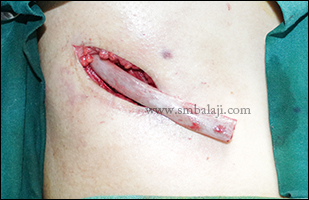
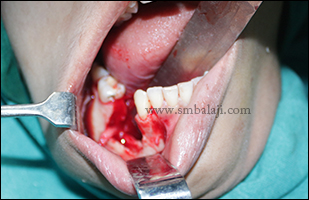
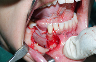
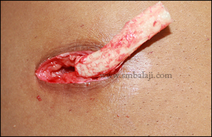
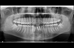
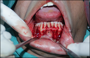
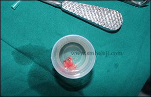
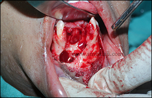
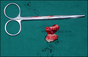
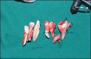
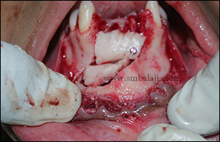
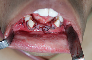
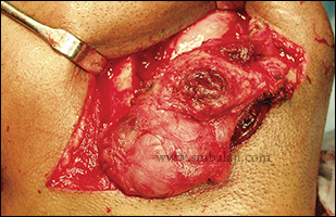
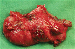
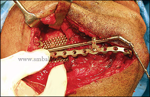
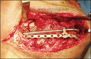
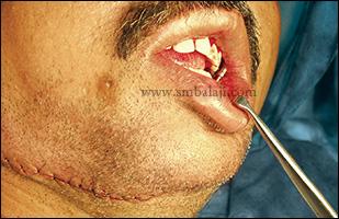
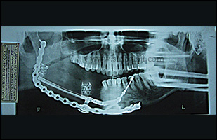
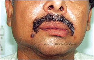
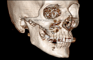
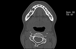
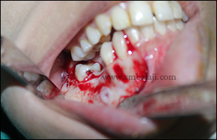
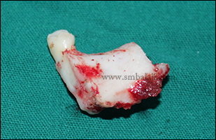
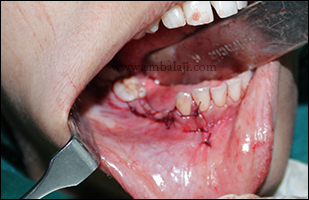
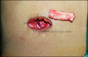
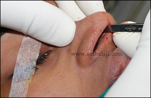
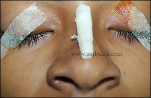
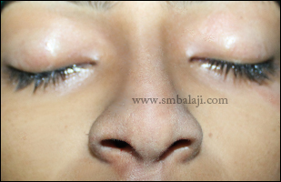
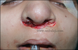
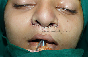
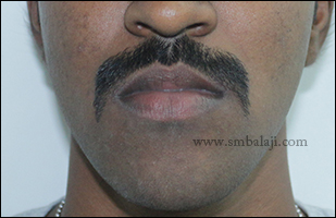
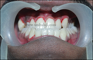
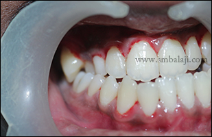
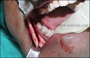
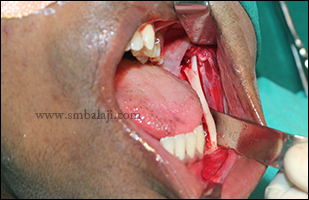
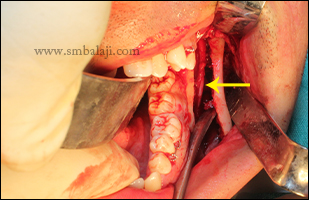
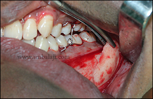
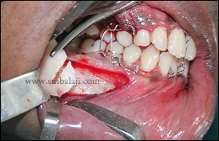
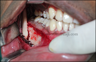
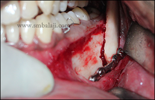
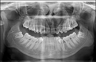
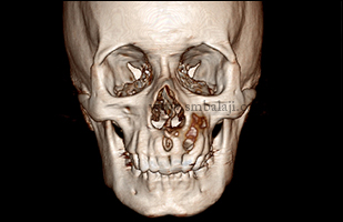
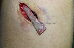
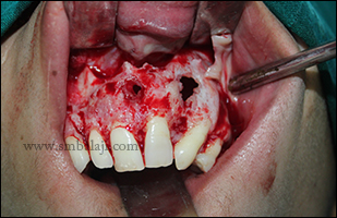
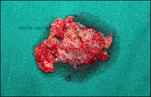
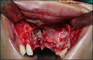
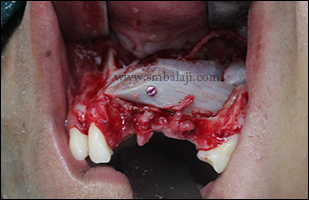
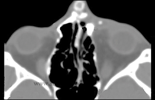
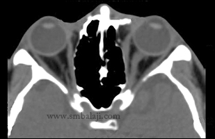
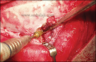
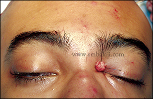
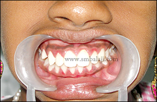
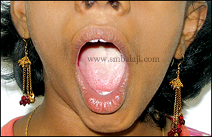
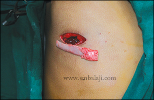
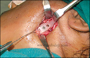
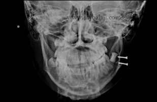
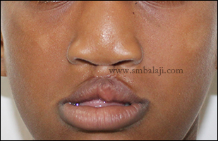
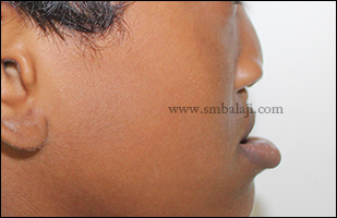
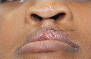
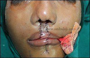
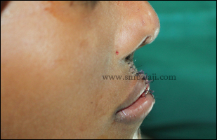
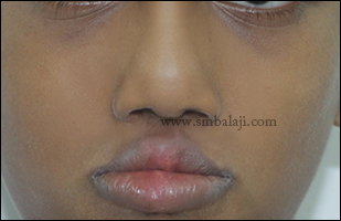
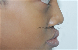
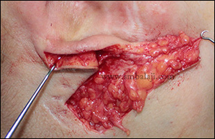
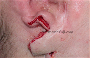
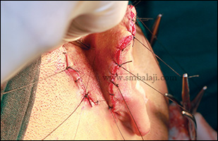
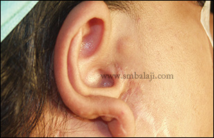
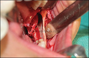
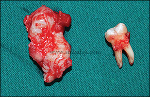
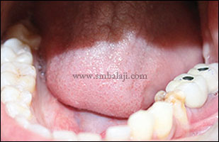
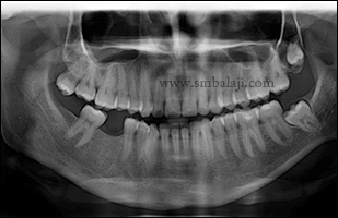
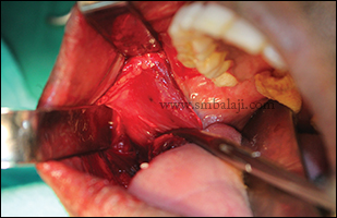
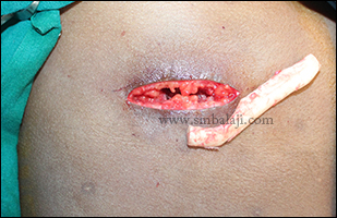
After thorough clinical, radiological and histopathological examination, Maxillofacial Surgeon Dr. S. M. Balaji diagnosed it as ameloblastoma involving the entire anterior segment of mandible. Surgery was planned to remove the lesion and reconstruct the affected jaw portion using costochondral rib graft in a single stage. Intraorally, complete removal of the lesion along with the involved teeth with sufficient clearance of the bone was given. Costochondral rib graft was fixed to reconstruct the mandibular defect. Incision was closed in layers. Patient feels very happy to have both removal and reconstruction in a single surgery.
After thorough clinical, radiological and histopathological examination, Maxillofacial Surgeon Dr. S. M. Balaji diagnosed it as ameloblastoma involving the entire anterior segment of mandible. Surgery was planned to remove the lesion and reconstruct the affected jaw portion using costochondral rib graft in a single stage. Intraorally, complete removal of the lesion along with the involved teeth with sufficient clearance of the bone was given. Costochondral rib graft was fixed to reconstruct the mandibular defect. Incision was closed in layers. Patient feels very happy to have both removal and reconstruction in a single surgery.


 RSS Feed